




Abstract
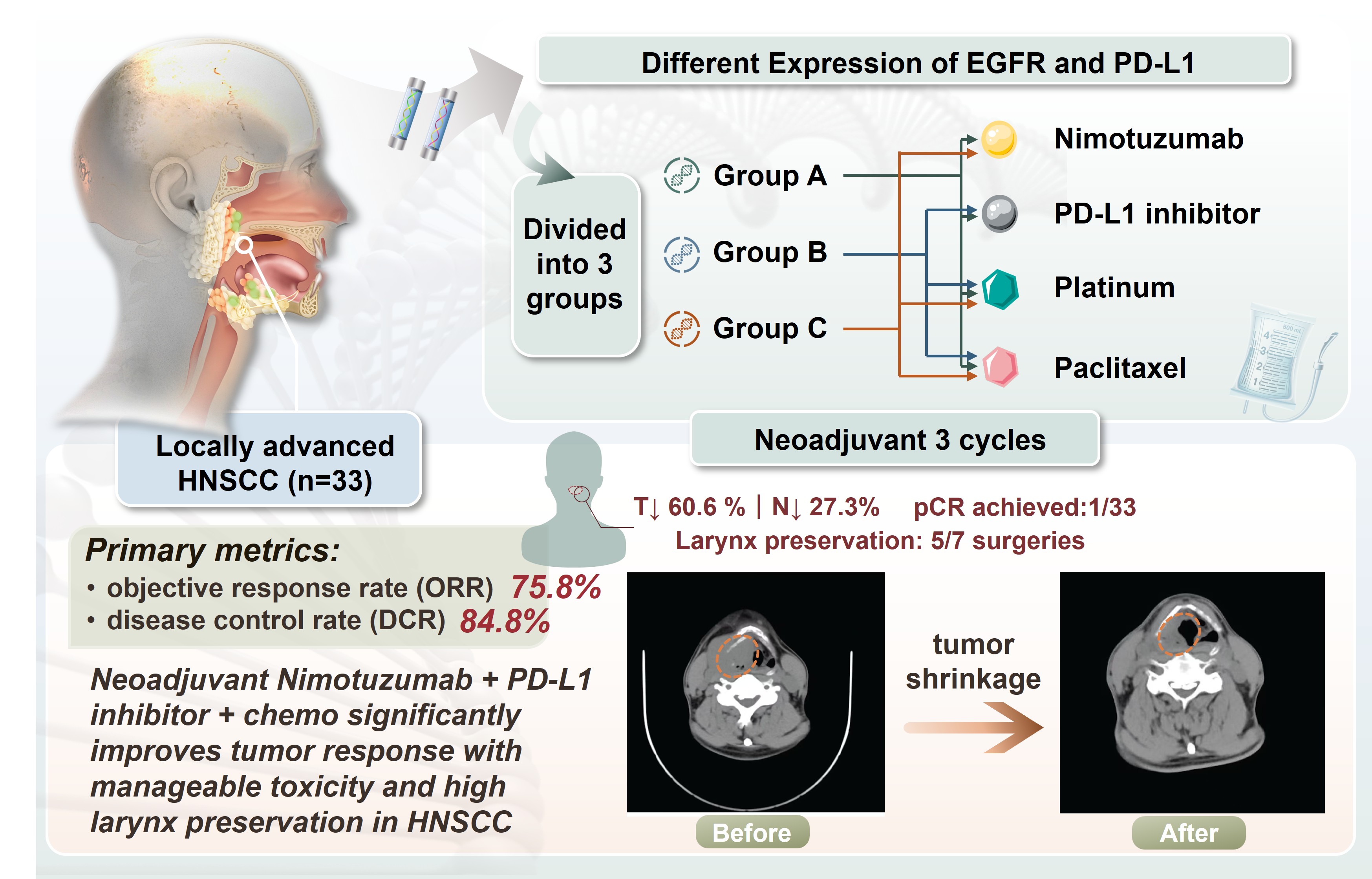
Objective: Our study aimed to assess the effectiveness and safety of neoadjuvant therapy involving the immune checkpoint inhibitor Nimotuzumab and programmed cell death ligand 1 (PD-L1) inhibitors, along with platinum and paclitaxel chemotherapy in individuals diagnosed with the head and neck squamous cell carcinoma (HNSCC).
Methods: Over the course of 2024 and 2025, a cohort of 33 patients with locally advanced HNSCC admitted to our institution was included in the investigation. Stratification into three groups was based on the expression levels of epidermal growth factor receptor (EGFR) and PD-L1 in tumour tissues. The patients were treated with Nimotuzumab and PD-L1 inhibitors along with platinum and paclitaxel chemotherapy for three cycles. After three cycles of neoadjuvant therapy, some patients were given surgical treatment. The primary metrics for evaluating treatment success were the objective response rate (ORR) and the disease control rate (DCR). Secondary endpoints included pathological complete response rate, laryngeal preservation rate, and incidence of chemotherapy-related toxicities.
Results: The study results revealed promising tumour response among the 33 patients, with an objective response rate of 75.8%. One patient achieved a complete radiographic response. Twenty patients achieved radiologic response, with T-stage downstaging observed in 60.6% and N-stage downstaging in 27.3% of these cases. Seven patients underwent surgery following neoadjuvant therapy, with five of these patients successfully undergoing larynx-preserving hypopharyngeal cancer radical resections. Regarding adverse events, 21.2% of patients experienced leukopenia; 42.4% experienced anemia; 3.0%, hepatic impairment; and 9.1%, gastrointestinal reactions.
Conclusions: Neoadjuvant combination therapy significantly enhances tumour response rates in HNSCC, with most patients with laryngeal cancer retaining laryngeal function. Adverse reactions remain clinically manageable, and the majority of patients tolerate the treatment well.
Keywords: Head and neck squamous cell carcinoma, Neoadjuvant therapy, Targeted therapy, Immunotherapy
Introduction
Head and neck malignancies represent a diverse group of cancers arising in various regions of the head and neck, excluding the eyes, brain, ears, esophagus, and thyroid[1, 2]. These cancers are the sixth most prevalent malignancy globally, with a particularly significant impact in China, and more than 500000 patients worldwide have been diagnosed with head and neck squamous cell carcinoma (HNSCC)[2, 3]. HNSCC is particularly aggressive, exhibiting a poor prognosis and a substantial societal and healthcare burden[4]. The incidence of HNSCC has increased by 25% over the past decade, with a five-year survival rate ranging between 40% and 50%[5, 6]. The majority of patients are diagnosed at locally advanced stages (III-IVB), for whom the standard of care is concurrent chemoradiotherapy using platinum-based agents, primarily cisplatin[7, 8]. However, this regimen has failed to significantly enhance long-term survival rates, and the adverse effects of platinum drugs, such as nephrotoxicity, neurotoxicity, and bone marrow suppression, often impair treatment compliance[4, 9]. Given these challenges, novel treatment approaches have emerged, particularly neoadjuvant therapy prior to surgery[8]. Molecular targeted therapy and immunotherapy have revolutionized the management of HNSCC, offering promising avenues for breakthrough progress in treating these malignancies[8]. HNSCC is generally responsive to chemotherapy, making induction chemotherapy an effective strategy to reduce distant metastasis rates and improve long-term survival. Preoperative induction chemotherapy not only mitigates the risk of distant metastasis but also enhances the prognosis by shrinking the tumour volume, eradicating micrometastases, and lowering clinical staging, which facilitates subsequent curative surgery and/or radiotherapy[10]. As an adjunctive preoperative intervention, neoadjuvant chemotherapy enables patients to undergo surgery or radiotherapy. Recent studies have demonstrated that neoadjuvant chemotherapy offers distinct advantages in terms of enhancing pathological response rates and improving survival outcomes[6]. When combined with targeted therapies or immune checkpoint inhibitors, synergistic therapeutic effects can be achieved[10]. Epidermal growth factor receptor (EGFR) overexpression is common in most HNSCC cases and is closely associated with tumour proliferation, invasion, and resistance mechanisms, making it a key therapeutic target[3, 11, 12]. Nimotuzumab, a humanized monoclonal antibody targeting EGFR, offers distinct mechanisms of action compared to other EGFR therapies and has demonstrated a relatively high safety profile with robust clinical trial data[3]. The 2024 edition of the CSCO Head and Neck Cancer Diagnosis and Treatment Guidelines recommends nimotuzumab as a treatment option for locally advanced HNSCC. Additionally, a subset of HNSCC patients exhibits PD-L1 expression in tumour cells. In these patients, PD-L1 inhibitors may demonstrate efficacy against tumours expressing this marker, offering a potential therapeutic benefit[13]. For PD-L1-positive patients, corresponding inhibitor therapies may provide a promising treatment option[5]. Overall, the evolving landscape of HNSCC treatment highlights the importance of personalized therapeutic strategies incorporating neoadjuvant chemotherapy, targeted therapies, and immunotherapies to improve patient outcomes[1].
Materials and Methods
General InformationBetween January 2024 and December 2025, 33 patients diagnosed with locally advanced head and neck squamous cell carcinoma were enrolled. The age range of the patients varied widely, spanning from 34 to 81 years old, with a marked gender disparity, as 31 patients (93.9%) were male and only 2 (6.1%) were female (Table 1). All patients received histopathological confirmation of squamous cell carcinoma before initiating neoadjuvant therapy. In terms of anatomical location, the most prevalent cancer type was hypopharyngeal carcinoma, accounting for 22 cases (66.7%). Tonsillar carcinoma was diagnosed in 3 patients (9.1%), laryngeal carcinoma in 3 patients (9.1%), oropharyngeal carcinoma in 3 patients (9.1%), and tongue carcinoma in 2 patients (6.1%) (Table 2). Regarding staging, the majority of patients were classified as Stage IV, with 28 cases (84.8%), while Stage III was observed in 5 cases (15.2%). In terms of T staging, 4 patients (12.1%) were in T1 stage, 17 patients (51.5%) were in T2 stage, and 12 patients (36.4%) were in T3 stage.For N staging, 2 patients (6.1%) were categorized as N0 stage, 3 patients (9.1%) as N1 stage, 27 patients (81.8%) as N2 stage, and 1 patient (3.0%) as N3 stage (Table 1). This diverse group of patients served as the foundation for our study, allowing for a comprehensive investigation into the efficacy and safety of neoadjuvant therapy in the treatment of locally advanced head and neck squamous cell carcinoma.
Table 1. Gender, age, clinical stage, T stage, and N stage of the 33 patients.
| Classification | Group A | Group B | Group C | Summation(%) | |
|---|---|---|---|---|---|
| Gender | Man | 8 | 8 | 15 | 31(93.9) |
| Woman | 0 | 2 | 0 | 2(6.1) | |
| Age | ≤57y | 2 | 4 | 7 | 13(39.4) |
| >57y | 6 | 6 | 8 | 20(60.6) | |
| Clinical staging | III | 0 | 2 | 3 | 5(15.2) |
| IV | 8 | 8 | 12 | 28(84.8) | |
| T stage | T1 | 0 | 2 | 2 | 4(12.1) |
| T2 | 5 | 4 | 8 | 17(51.5) | |
| T3 | 3 | 4 | 5 | 12(36.4) | |
| N stage | N0 | 0 | 0 | 2 | 2(6.1) |
| N1 | 0 | 2 | 1 | 3(9.1) | |
| N2 | 8 | 8 | 11 | 27(81.8) | |
| N3 | 0 | 0 | 1 | 1(3.0) | |
Table 2. Number and percentage of the 33 patients with different primary lesions.
| Classification | Group A | Group B | Group C | Summation(%) |
|---|---|---|---|---|
| Hypopharynx | 5 | 6 | 11 | 22(66.7) |
| Tonsil | 1 | 0 | 2 | 3(9.1) |
| Larynx | 0 | 2 | 1 | 3(9.1) |
| Oropharynx | 2 | 0 | 1 | 3(9.1) |
| Tongue | 0 | 2 | 0 | 2(6.1) |
This study was approved by the ethics committee. As this research is a retrospective study, the ethics committee waived the requirement for obtaining informed consent. This study complies with the guidelines of the Helsinki Declaration. The study included patients aged 18 years and older with histologically/cytologically and radiologically confirmed, locally advanced (stage III-IV) or recurrent/metastatic squamous cell carcinoma of the head and neck, primarily affecting the oral cavity, oropharynx, hypopharynx, or larynx. Patients were deemed inoperable due to refusal or medical contraindications to surgery/anaesthesia. They had normal bone marrow, hepatic, and renal function, with an expected survival of at least 6 months. Participants had no prior anti-PD-L1 therapy, radiotherapy, or contraindications to chemoradiotherapy.
Exclusion CriteriaExclusion criteria included:(1) history of anti-EGFR therapy,(2) disease progression during or after prior platinum-based therapy,(3) uncontrolled systemic conditions such as active infections or HIV,(4) severe cardiac insufficiency,(5) severe pulmonary disease or history of interstitial lung disease,(6) known allergies to monoclonal antibodies (especially murine or humanised) or chemotherapeutic agents (paclitaxel, platinum-based drugs),(7) pregnant or lactating women, as well as fertile patients who refused contraception.
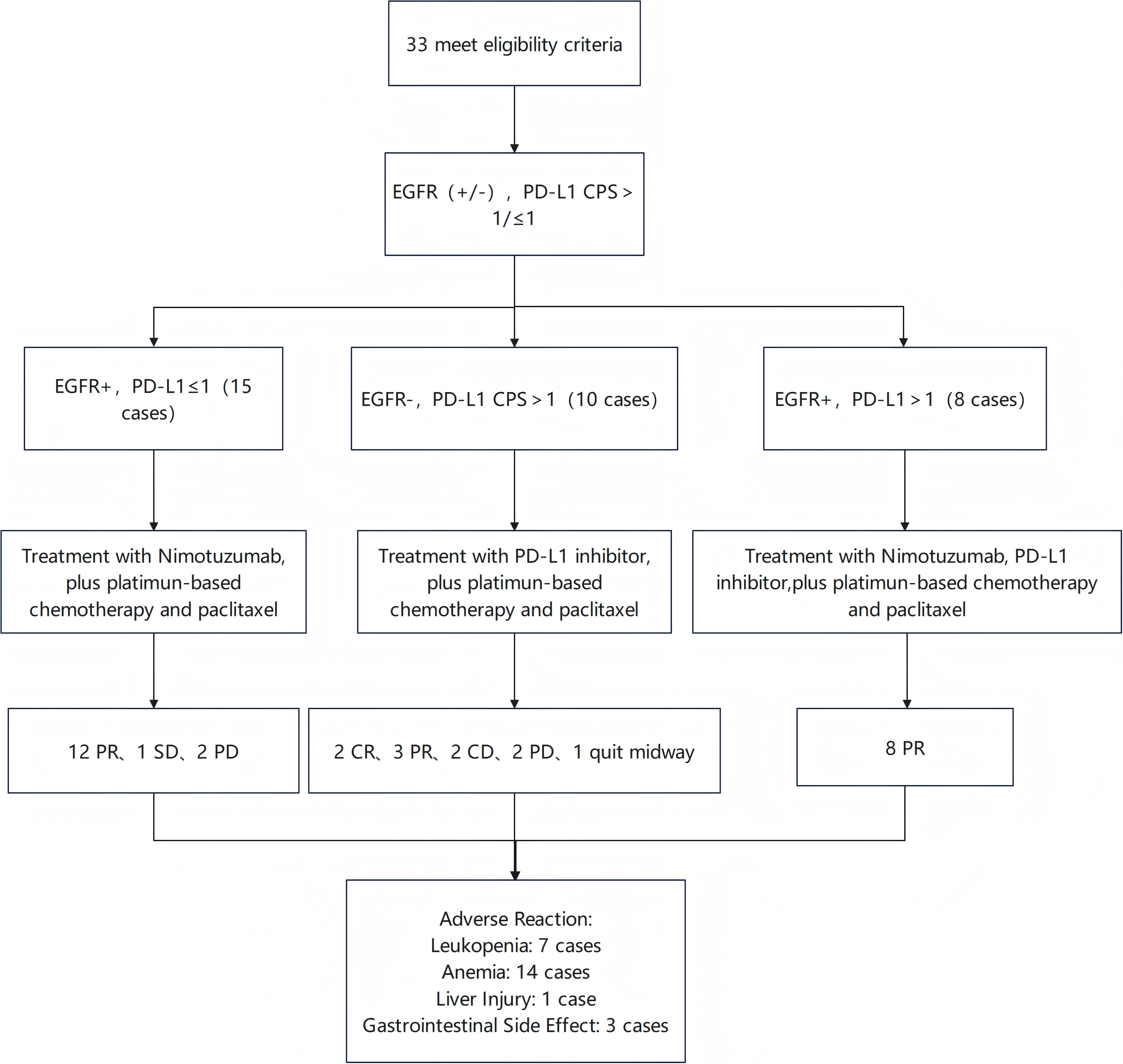
Treatment ProtocolThe flowchart of this research is shown in Figure 1. The detection of EGFR and PD-L1 CPS score is a standardized pathological process, primarily relying on immunohistochemistry. The test results in this study were certified by the Department of Pathology at the First Affiliated Hospital of Anhui Medical University. After obtaining samples through biopsy, sections were prepared for immunohistochemical staining using the VENTANA platform. Pathologists calculated the CPS value based on the counting results. A CPS greater than 1 indicates consideration for PD-L1 inhibitor therapy, and a higher CPS value suggests a greater likelihood of benefiting from immunotherapy. Based on EGFR and PD-L1 expression levels, tumour cell count, and CPS results, patients were randomized into three groups, A, B, and C. Patients in group A received a combination of Nimotuzumab, a PD-L1 inhibitor, platinum-based chemotherapy, and paclitaxel. Patients in group B received a combination of a PD-L1 inhibitor, platinum-based chemotherapy, and paclitaxel. Patients in group C received a combination of Nimotuzumab, platinum-based chemotherapy, and paclitaxel, all administered for three cycles. The primary endpoints were objective response rate (ORR) and disease control rate (DCR). Secondary endpoints included pathological complete response rate, larynx preservation rate, and chemotherapy-related toxicity.
Figure 1. Flowchart of the study protocol.
After three cycles of neoadjuvant therapy, tumour response was evaluated using the Response Evaluation Criteria in Solid Tumours (RECIST 1.1). Specific imaging classifications were determined as follows, a complete response (CR) was defined as the disappearance of all target lesions for at least 4 weeks, a partial response (PR) was defined as a ≥30% reduction in the sum of the longest diameters of target lesions compared to baseline,maintained for at least 4 weeks, progressive disease (PD) was defined as either an increase in target lesion size not meeting PR criteria or the appearance of new target lesions, and stable disease (SD) was defined as lesions not meeting PR or PD criteria. The objective response rate served as an indicator of the treatment's antitumour activity, while the disease control rate reflected its ability to halt recent disease progression. For patients who underwent surgical intervention, postoperative pathological assessments of the primary lesion were conducted to determine the pathological complete response rate. For those who underwent curative surgery, laryngeal function preservation rates were calculated. Furthermore,the study meticulously documented any adverse reactions throughout the treatment period,encompassing gastrointestinal reactions such as nausea and vomiting, myelosuppression manifesting as leucopenia and anaemia, and hepatic dysfunction. These comprehensive evaluations allowed for a detailed assessment of both the efficacy and safety profile of the neoadjuvant therapy regimen employed in this study.
Statistical AnalysisData from the research study were analyzed using SPSS 27 statistical software, with a focus on employing descriptive statistical methods. The study included the calculation of various rates for different variables. For instance, the tumour response rate was calculated by dividing the sum of complete response cases and partial response cases by the total number of treated cases, then multiplying by 100%. The larynx preservation rate was determined by dividing the number of partial laryngectomies by the total number of surgical cases, multiplying by 100%. The pathological complete response rate was calculated by dividing the cases with no detectable tumour in postoperative pathology by the total treated cases, then multiplying by 100%. Additionally, the adverse reaction rate was determined by dividing the number of adverse reaction episodes by the total treatment episodes, then multiplying by 100%. All statistical results were presented with one decimal place for accuracy. The incidence rate was calculated as: number of adverse events/total number of treated cases × 100%. All statistical results were rounded to one decimal place.
Results
Tumour Response Rate and Disease Control RateFollowing completion of neoadjuvant therapy, all patients underwent efficacy assessment using imaging modalities consistent with baseline. Of these, 32 patients successfully completed all three treatment cycles, with the exception of one patient who was lost to follow-up after the first course of neoadjuvant therapy. Efficacy evaluation showed that the disease control rate for the entire cohort was 84.8% (28/33) and the objective response rate was 75.8% (Table 3). Specifically, two patients achieved complete response after completing three cycles, 23 patients attained partial response, three patients demonstrated stable disease, and four patients experienced disease progression. Among HNSCC patients completing three cycles, 20 (60.6%) achieved radiologic response: 20 (60.6%) achieved T-stage downstaging and 9 (27.3%) achieved N-stage downstaging.
Table 3. Evaluation of the efficacy of neoadjuvant treatment in 33 patients with HNSCC.
| Group(n) | CR | PR | SD | PD | Quit midway | ORR(%) | DCR(%) |
|---|---|---|---|---|---|---|---|
| A(n=8) | 0 | 8 | 0 | 0 | 0 | 100 | 100 |
| B(n=10) | 2 | 3 | 2 | 2 | 1* | 50 | 70 |
| C(n=15) | 0 | 12 | 1 | 2 | 0 | 80 | 86.7 |
| Summation(n=33) | 2 | 23 | 3 | 4 | 1 | 75.8 | 84.8 |
*One patient was lost to follow-up after only one cycle of neoadjuvant treatment.
After one patient was lost to follow-up, another patient successfully achieved complete radiologic response after three cycles of neoadjuvant therapy. Among the patients who completed the three cycles, seven underwent surgery. Five patients underwent radical surgery for hypopharyngeal cancer with successful preservation of laryngeal function, achieving a laryngeal preservation rate of 100%. Postoperative pathological reports for all surgical patients showed that one patient achieved pathological complete response of the primary lesion, accounting for 3.03% of all enrolled patients. Two patients with tonsillar squamous cell carcinoma underwent surgery: one had radical neck dissection and the other had extended resection of the tonsillar carcinoma combined with radical neck dissection, with postoperative pathology indicating no cancer cells in the primary tonsillar tissue.
Adverse Reaction IncidenceAdverse reactions are common during neoadjuvant therapy, with bone marrow suppression, gastrointestinal issues, and hepatic dysfunction being the most frequent. Out of 33 patients, 21.2% developed leukopenia, mainly Grade I-II reactions, while 42.4% had anaemia, with one patient experiencing Grade III reactions (Table 4). A single individual (3.0%) suffered from severe Grade IV hepatotoxicity. Gastrointestinal reactions like nausea and vomiting affected 9.1% of patients, all classified as Grade I-II reactions. Interestingly, no cases of thrombocytopenia were reported post-treatment. Despite these challenges, patients continued their therapy journey, showcasing resilience and determination throughout their treatment.
Table 4. Adverse reactions to neoadjuvant therapy in 33 patients with HNSCC
| Adverse Reaction | Group A | Group B | Group C | Percentage(%) | Adverse Reaction Grading | |||
|---|---|---|---|---|---|---|---|---|
| I | II | III | IV | |||||
| Leukopenia | 2 | 1 | 4 | 21.2 | 5 | 2 | 0 | 0 |
| Anemia | 4 | 2 | 8 | 42.4 | 10 | 3 | 1 | 0 |
| Thrombocytopenia | 0 | 0 | 0 | 0 | 0 | 0 | 0 | 0 |
| Hepatotoxicity | 0 | 1 | 0 | 3.0 | 0 | 0 | 0 | 1 |
| Gastrointestinal Reaction | 3 | 0 | 0 | 9.1 | 3 | 0 | 0 | 0 |
Discussion
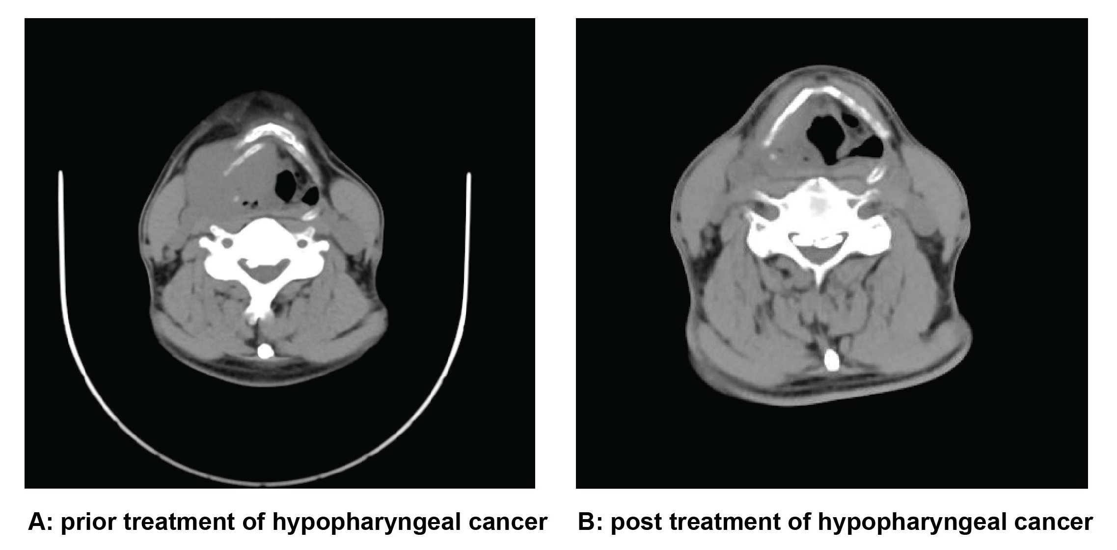
HNSCC is often diagnosed at an advanced stage, leading to poor five-year survival rates despite current treatment strategies. The standard treatment approach involves a combination of cisplatin and paclitaxel-based chemotherapy with surgery, which often results in complications such as difficulty swallowing and speaking, negatively impacting the quality of life for patients post-surgery. However, recent studies have shown promising outcomes with the use of immunotherapy or targeted agents in conjunction with neoadjuvant chemotherapy, which may lead to better tumour shrinkage and increase the feasibility of radical surgery [10]. This innovative approach not only helps preserve organs but also improves the long-term quality of life for HNSCC patients. For locally advanced HNSCC patients, preoperative neoadjuvant therapy is becoming a strategy that can significantly improve prognosis. Its main advantages are as follows: shrinking the tumor, improving surgical outcomes, enhancing pathological remission, improving survival prognosis, early elimination of micrometastases, reducing the risk of recurrence, assessing drug sensitivity, and guiding subsequent treatment. The results of this study demonstrated that enrolled patients achieved an overall ORR of 75.8% and a DCR of 84.8% following neoadjuvant therapy, with a 6.1% incidence of Grade III-IV treatment-related adverse events. As shown in Figure 2, the primary tumour exhibited significant shrinkage after three cycles of neoadjuvant therapy. Based on the expression of EGFR and PD-L1 biomarkers, the neoadjuvant regimen incorporating Nimotuzumab, PD-L1 inhibitors and chemotherapy demonstrated promising antitumour activity with an overall manageable safety profile. This study demonstrates that the combination of immunotherapy, targeted therapy, and chemotherapy as a neoadjuvant regimen offers a promising treatment option for HNSCC. In fact, the 2024 CSCO guidelines have officially recommended the use of Nimotuzumab, a humanized anti-EGFR monoclonal antibody, in combination with concurrent chemoradiotherapy for patients with locally advanced nasopharyngeal carcinoma (Category 1A evidence, Class I recommendation).
Figure 2. Comparative imaging of a patient with hypopharyngeal cancer before and after neoadjuvant therapy.
On the other hand, in patients with high PD-L1 expression, combining PD-L1 inhibitors with chemotherapy has consistently shown positive results, supporting the theories of tumour immunology[5]. The findings of this study demonstrated that the ORR reached 50% in patients treated with PD-L1 inhibitors combined with chemotherapy, including two cases achieving CR. PD-L1 expression suggests a pre-activated immune state in the tumour microenvironment, but it also indicates the presence of immune checkpoint-mediated immunosuppression. PD-L1 inhibitors can remove this “braking” signal and reinvigorate T cell-mediated tumour killing. Furthermore, the combination of PD-L1 inhibitors with chemotherapy can induce immunogenic cell death, potentially creating a “1+1 > 2” synergistic anti-tumour effect[9, 15]. The evidence provided clearly demonstrates the effectiveness of immune checkpoint inhibitors in treating head and neck cancer[16]. Particularly in patients with high levels of PD-L1 and EGFR, using both types of treatment concurrently produces better outcomes that achieve a high ORR. The success of treatment for these individuals may depend on the balance between EGFR activity and immune suppression in their tumour microenvironment. Looking ahead, combining targeted therapy, immunotherapy, and chemotherapy as neoadjuvant treatment for advanced cancers may offer promising advances. However, the key remains conducting a thorough evaluation of various factors such as tumour characteristics, staging, genetic markers, and overall health to tailor treatment plans to each individual patient's unique needs.
In this study, the spectrum of adverse reactions observed remained within manageable limits, with tumour-related complications primarily being anaemia. The most prevalent adverse reaction noted was bone marrow suppression, predominantly Grade I-II toxicity. The adverse effects of neoadjuvant therapy are within a controllable range, and proactive prevention, close monitoring, and early intervention measures are crucial in effectively managing these adverse reactions. Therefore, healthcare professionals should be vigilant in ensuring the well-being of patients undergoing treatment, making careful consideration of potential complications and taking prompt action when necessary.
It is important to note that this study has limitations that need to be taken into consideration. Firstly, it was conducted in a single center with a small sample size, which may limit the generalizability of the results. The sample size in this study is relatively small, which may affect the robustness and reliability of the research findings. Additionally, the lack of validation from large-scale, randomised controlled clinical trials calls for caution when interpreting the findings. The three-cycle treatment duration was relatively short, which may have prevented the observation of long-term benefits of neoadjuvant combination therapy. Moreover, potential delayed immune-related adverse events may not have been detected due to the limited follow-up period. Moving forward, future studies should consider incorporating factors such as tumour mutation burden, HPV status, and refined tumour microenvironment subtyping to enhance the accuracy of efficacy prediction models. The new neoadjuvant treatment approach combining immunotherapy, targeted therapy and chemotherapy will be the main direction for the future treatment of HNSCC.
Conclusion
Histological analysis of EGFR and PD-L1 expression in HNSCC patients helped in categorizing them into different treatment groups. Nimotuzumab, PD-L1 inhibitors, paclitaxel, and platinum-based chemotherapy have a good therapeutic effect on patients with locally advanced HNSCC and can significantly reduce the size of the tumor. Following three cycles of treatment, promising efficacy was observed in these patients, accompanied by manageable adverse reactions. This personalized approach demonstrates the potential for tailored precision treatment in HNSCC cases. It sets the stage for broader clinical trials that aim to enhance the overall survival and quality of life for individuals battling HNSCC by employing the most effective treatment regimens.
Abbreviations
CR - complete response; DCR - disease control rate; EGFR - epi-dermal growth factor receptor; HNSCC - head and neck squa-mous cell carcinoma; ORR - objective tumor response rate; PD - progressive disease; PD-L1 - programmed cell death ligand 1; PR - partial response; SD - stable disease
Declarations
Author Contributions
Nuo Chen: Investigation, Data curation, Formal analysis, Writing - original draft; Dong Wang: Conceptualization, Methodology, Supervision, Writing - review & editing; Chaobing Gao: Conceptualization, Supervision, Project administration.
Acknowledgements
We thank the medical staff and patients who participated in this study.
Funding information
Not Applicable.
Ethics Approval and Consent to Participate
This study was approved by the Ethics Committee of the First Affiliated Hospital of Anhui Medical University (Approval No. PJ 2025-08-72). Written informed consent was obtained from all participants.
Competing Interests
The authors declare that they have no existing or potential commercial or financial relationships that could create a conflict of interest at the time of conducting this study.
Data availability
All data generated or analyzed during this study are included in this published article.
References
 Figures
 References
 Peer
Peer Information
Figure 1. Flowchart of the study protocol.
Figure 2. Comparative imaging of a patient with hypopharyngeal cancer before and after neoadjuvant therapy.
Peer-review Terminology
Identity transparency: Single anonymized
Reviewer interacts with: Editor
Details
This is an open access article under the terms of the Creative Commons Attribution License(http://creativecommons.org/licenses/by/4.0/), which permits use, distribution and reproduction in any medium, provided the original work is properly cited.
Publication History
Received 2025-10-22
Accepted 2025-11-14
Published 2025-12-14