




Abstract
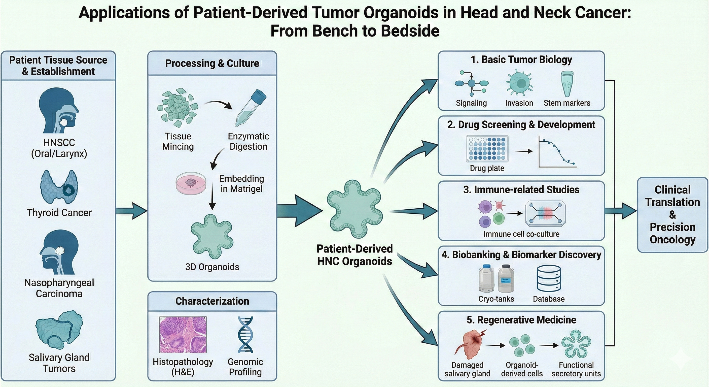
Head and neck cancers (HNC) constitute a highly heterogeneous group of malignancies with complex pathogenesis; effective clinical management remains challenging. Organoids, an emerging three-dimensional culture system, recapitulate key structural and functional features of the tissues of origin and offer distinctive advantages for studying head and neck cancer. Structured around four major head and neck cancer entities (head and neck squamous cell carcinoma, thyroid cancer, nasopharyngeal carcinoma, and malignant salivary gland tumors), this Review systematically outlines approaches for constructing head and neck cancer organoids, summarises common modelling methods and authentication strategies, and defines an integrated pipeline spanning acquisition, culture, validation and application. At the application level, we review five areas: basic tumor biology; drug screening and new drug development; immune-related studies; biobanking and biomarker discovery; and regenerative medicine with functional reconstruction. The goal is to inform subsequent research and clinical translation, and to highlight the broad prospects of this approach for addressing heterogeneity, therapy resistance and recurrence in head and neck cancer.
Keywords: Head and neck cancers, Patient-derived tumor organoids, Drug screening, Biomarker discovery, Regenerative medicine
Introduction
Head and neck cancers (HNC) account for roughly one tenth of all malignancies worldwide, with an estimated 600,000–900,000 new cases annually[1]. In clinical practice they are grouped into head and neck squamous cell carcinoma (HNSCC), salivary gland carcinoma (SGC) and thyroid cancer (TC)[2]. HNSCC is the predominant subtype, with major risk factors including heavy alcohol use, tobacco exposure, and human papillomavirus infection[3, 4]. In 2024, there were about 891,000 new HNC cases globally; nearly 90% were squamous cell carcinomas, and ~60% presented at a locally advanced stage, contributing to poor outcomes[5].
In the precision medicine era, tools that support individualised therapy selection are urgently needed. While 2D-cancer cell lines are widely used for drug screening, they fail to realistically predict clinical responses[6]. Similarly, animal models and patient-derived xenografts (PDX) are constrained by species differences, high costs, and the tendency for murine stroma to replace the human niche over time, which confounds pharmacological assessments[7]. These limitations hinder timely and reproducible response prediction within clinical timelines.
Three-dimensional (3D) culture offers a superior alternative to these conventional models. Tumor spheroids were the earliest 3D systems, but because they usually originate from single cell lines and have simplified architecture, they do not faithfully reproduce native tissue structure or function[8]. Unlike tumor spheroids, which typically originate from single cell lines and lack structural complexity, organoids are self-organising models that reconstruct tissue-like cellular composition and spatial organisation[9]. Since the pioneering establishment of intestinal organoids by Sato et al. , culture conditions have been optimized for various tissues, including brain, liver, and lung[10, 11]. Crucially, unlike cell lines, patient-derived organoids (PDOs) maintain primary genomic alterations and functional heterogeneity, providing a high-fidelity platform for modelling the tumor microenvironment and drug screening[10, 11].
In head and neck oncology, PDOs can be established from surgical specimens or core biopsies within weeks[12]. They preserve key driver alterations and histo-architectural features, enabling parallel testing of radiochemotherapy regimens and co-culture with autologous immune cells[13, 14]. Systematic biobanking of PDOs further supports longitudinal studies and cross-centre benchmarking. However, challenges such as matrix variability, incomplete immune reconstitution, and the need for standardisation remain.
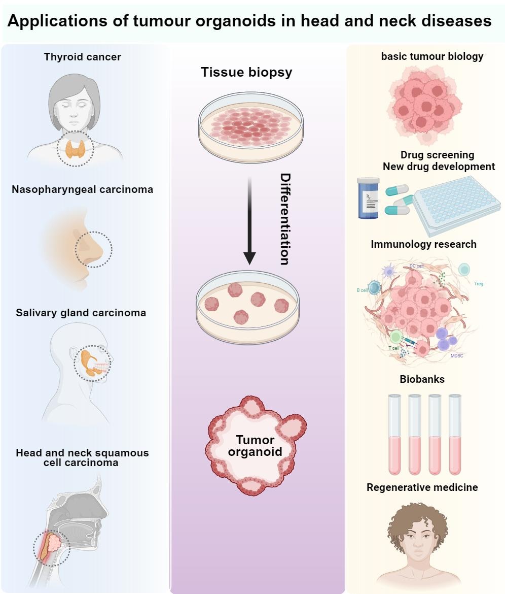
This Review focuses on organoid construction and therapeutic-response prediction for four major head and neck entities: HNSCC, TC, NPC, and SGC. We outline practical workflows for establishment and authentication, and evaluate applications across basic biology, drug screening, immune-related studies, biomarker discovery, and regenerative medicine. Finally, we discuss integration with emerging technologies like organoids-on-chips and multi-omics to address current limitations and advance clinical translation (Figure 1).
Figure 1. Schematic of the sources and applications of tumour organ-oids in head and neck diseases (Created with bioRender.com).
Methods for establishing head and neck cancer organoids
The development of head and neck tumor organoid spheroids proceeds through several critical steps. The foremost is selecting an appropriate tissue or organ source; the next is choosing a suitable culture scaffold—whether derived from natural matrices or formulated from synthetic materials—which is pivotal for growth and functional fidelity.
Tissue selection and pre-analytical handlingSpecimen acquisition and pre-analytics set the upper limit for organoid establishment. Resections, endoscopic or fine-needle biopsies, and samples from recurrent or metastatic sites are acceptable sources[15, 16]. Cold ischemia time should be minimised, and pathology review, tumor cellularity estimation, and documentation of viral status should be completed. Disease-specific points apply. HNSCC typically yields ample cells, but fibroblast overgrowth must be controlled. In 2018, Tanaka et al. proposed the cancer tissue–originated spheroid method, processing tissue from 43 patients into small fragments rather than single cells; clusters formed spheroids within 24 hours and, after embedding, efficiently generated head and neck tumor organoids while reducing anoikis[17]. Nasopharyngeal carcinoma often provides limited tissue; gentle enzymatic digestion and rapid matrix embedding improve viability. Researchers established NPC organoids from patient tumors, authenticated them by morphology, histopathology and immunohistochemistry (Ki-67, CD133) together with EBER in situ hybridisation, and used them for drug testing[18]. Thyroid cancer specimens frequently contain colloid and thrombi; these should be removed by filtration before counting and seeding, and driver alterations such as BRAF, RET and NTRK should be recorded[18]. Ogundipe et al. developed patient-derived thyroid organoids with regenerative potential, showing lineage-specific cells that self-renew and differentiate into functional thyroid tissue[19]. Salivary gland tumors are slow-growing and stroma-rich, often requiring extended primary culture[20]; when needed, PDX-derived organoids can supplement cell yield for downstream assays. Researchers have established mouse and human salivary gland organoids that can be maintained long term, stably express acinar, myoepithelial and ductal lineage markers[21], and display secretory function in response to neurotransmitter stimulation. Human organoids can be initiated from either basal or luminal cells; single-cell RNA sequencing confirms retention of cellular heterogeneity and glandular diversity[22]. Across indications, serial passaging may favour dominant clones and erode intratumor heterogeneity, reducing concordance with the parental tumor and affecting reproducibility[23, 24]. Build flows and culture conditions should therefore be optimised by histology to maintain stability over time. For entities in which biopsy is difficult, alternative sourcing from tumor cells isolated from blood or other body fluids can enable organoid generation[25–28].
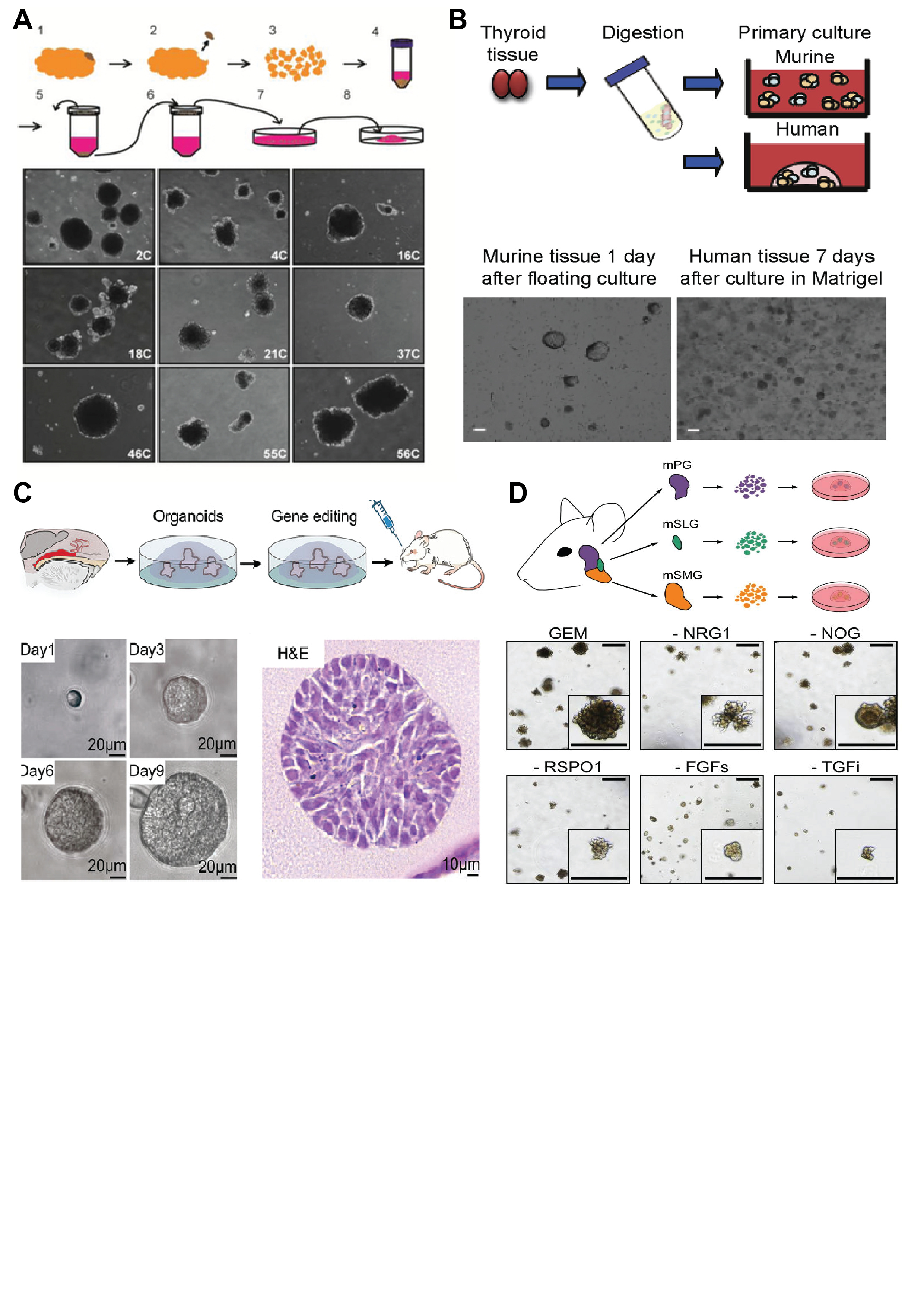
Construction methodsMost head and neck PDOs workflows share a common backbone, with differences confined to a few key steps. Following the scheme summarised by Kijima and Karakasheva[28], fresh tumor tissue is washed, mechanically minced and gently digested to a single-cell suspension or small clusters, then homogeneously mixed with Matrigel for embedding and expanded in defined media[29]. Medium formulations vary across studies, most notably in the choice and dosing of Wnt/R-spondin supplements, EGF/FGF family factors and TGF-β inhibitors[22, 28, 30]. Tanaka et.al proposed the cancer tissue-originated spheroid (CTOS) approach: patient tumor specimens are gently processed into small tissue fragments rather than single cells, enabling cell clusters to self-assemble into spheroids within 24 hours, thereby improving modelling efficiency and effectively reducing anoikis[17] (Figure 2A). Recent work has adopted a gentle dissociation workflow to generate highly viable single-cell suspensions from PDOs, using brief incubation with 0.05% trypsin coupled with mild mechanical dissociation; this approach avoids the aggregation and loss of viability seen with higher enzyme concentrations or prolonged exposure, markedly increases single-cell yield, and yields preparations suitable for downstream drug screening and single-cell RNA sequencing[31]. Furthermore, after tissue procurement, Ogundipe et al. mechanically and enzymatically dissociated murine and human thyroid tissue to obtain dispersed single cells: murine cells formed spheroids when suspended in a defined thyroid growth medium (TGM), whereas human cells were seeded directly into Matrigel and, after matrix polymerization, overlaid with human TGM supplemented with Wnt and R-spondin1 (TGM+WR), yielding spheroids in ~7 days[19] (Figure 2B). For nasopharyngeal carcinoma, where specimens are small and fibrotic, a “first-day suspension” variant has been used. After enzymatic dissociation, cells are kept overnight under low-adhesion conditions to self-aggregate into microclusters, then seeded into three-dimensional culture the next day. Compared with direct seeding, early aggregation enhances initial outgrowth and passaging efficiency (Figure 2C), which is advantageous when input tissue is limited. In rare and heterogeneous salivary gland tumors, methodology has an even greater impact on take rates[32]. Lassche and colleagues were the first to systematically establish and characterise SGC PDOs, reporting an overall success rate of about 19%, markedly lower than in many other tumor types [33]. To compensate for limited cell yield and slow proliferation, Aizawa and co-workers complemented patient-derived PDOs and PDXs with PDX-derived organoids (PDXOs), covering three SGC subtypes and recapitulating parental histology across all three models[30]. Recent work has established a workflow for salivary gland organoids with long-term maintenance, which stably express acinar, myoepithelial and ductal lineage markers and exhibit neurotransmitter-evoked secretory responses, thereby providing a platform for salivary gland regeneration and precision oncology assessment[22] (Figure 2D). To reduce the need for surgery or repeat biopsies, several groups have turned to circulating tumor cells. Using the eSelect system, Lin and colleagues expanded circulating tumor cells (CTCs) ex vivo to build organoids that mirrored clinical drug responses, achieving an overall establishment rate of 92.5%, substantially higher than with conventional tissue workflows[34]. This strategy suits advanced or metastatic disease in which tissue is scarce but is constrained in early disease by low CTC burden.
Figure 2. (A) Establishment of organoids from HNC tissue, and HNC organoids recapitulate thecharacteristics of original tumor tissues. Copyright © 2018 Noriaki Tanaka. (B) Schematic representation of murine and human thyroid primary cell culture. Thyroid gland tissue was mechanically and enzymatically digested and resuspended in culture medium or seeded in Matrigel. Copyright © 2021 Vivian M.L. (C) Mouse thyroid cells (n=3) after mechanical-enzymatic dissociation cultured in TGM; spontaneous spheroids at 24 h. Representative bright-field and 72 h high-magnifica-tion images. (D) Workflow for constructing primary and orthotopic NPC models, time-course bright-field images showing a monoclonal organoid arising from a single cell and representative H&E image of normal nasopharyngeal organoids. Copyright © 2024 Xudong Wan. (E) Experimental schematic for murine salivary gland organoids: parotid (mPG), sublingual (mSLG), and submandibular (mSMG) amd mSMG organoids cultured in complete growth expansion medium (GEM) or GEM lacking individual factors, with growth monitored by brightfield microscopy. Copyright © 2022, Yoon.
Taken together, the PDO pipeline should be tailored to tumor type, sample size and study purpose. When biological fidelity and speed are priorities, CTOS or micro-organosphere approaches are advantageous. When high-throughput screening and uniform plating are required, single-cell initiation is preferable. For rare subtypes or microenvironment-dependent questions, integrating PDX/PDXO sources with media optimisation is recommended, while remaining alert to culture-induced rewiring of critical signalling dependencies. Given the diversity of establishment protocols, we have summarized the key differences in culture conditions, media supplements, and specific challenges for distinct HNC subtypes in Table 1.
Table 1. Comparison of key culture conditions and establishment strategies for HNC organoid subtypes.
| HNC Subtype | Tissue Processing Strategy | Key Media Supplements | Specific Challenges |
|---|---|---|---|
| HNSCC | Mechanical mincing + enzymatic digestion; CTOS method for minimizing anoikis | Wnt3a, R-spondin, EGF, Noggin, TGF-β inhibitors (e.g., A83-01) | Controlling fibroblast overgrowth; managing rapid keratinization. |
| Thyroid Cancer (TC) | Filtration to remove colloid/thrombi; single cell seeding for human tissue | TSH (Thyroid Stimulating Hormone), Wnt, R-spondin1 | Retaining follicle structure and colloid production capability. |
| Nasopharyngeal (NPC) | "First-day suspension" aggregation to form microclusters before embedding | Rho-kinase inhibitor (Y-27632) critical for initial survival | Limited tissue quantity from endoscopic biopsies; heavy fibrosis. |
| Salivary Gland (SGC) | Extended primary culture; PDX-derived organoids (PDXOs) as supplement | Neurotransmitters (for functional secretory assays); FGF7/FGF10 | Slow growth rate; high stromal content requiring specific digestion. |
Organoids combine the scalability of cancer cell lines with a close resemblance to native tissues, making them powerful for drug discovery and basic research. Their successful propagation depends on lineage-specific supplements and meticulous technique, and outcomes can vary across operators and laboratories even from the same donor sample[35]. Accordingly, robust characterization is essential to verify similarity to the tissue of origin and to decide whether a batch is fit for pharmacological testing. In this section, we outline two commonly used approaches for organoid characterization: genomic profiling and histopathological assessment.
Genomic authenticationGenomic authentication establishes lineage identity between organoids and their paired tumors and assesses retention of driver events and intratumoral heterogeneity. The first step is identity verification using short tandem repeat profiling or SNP-based fingerprinting to match organoids to the patient specimen and exclude cross-contamination or cell-line mix-ups. Driver and genome-wide alterations are then compared using targeted panels or whole-exome sequencing (WES) to capture common somatic single-nucleotide variants and small insertions or deletions, complemented by shallow whole-genome sequencing or copy-number analysis to evaluate chromosomal instability and large-scale gains or losses[36]. Additionally, disease-specific viral signals should be recorded and cross-checked, for example EBV DNA or transcripts including EBER in situ hybridisation and LMP1 in nasopharyngeal carcinoma, and HPV-related readouts for oropharyngeal cases[32]. Researchers analyzed paired samples from untreated tumors (parent tumor and matched organoid) by performing WES on first-generation organoids, and used multi-region sampling to control for intratumoral heterogeneity[37]. Studies have shown that genomic profiling of 35 organoid cultures (WES, n=30) identifies tumor-derived models: only those harbouring driver mutations in established tumor suppressors or oncogenes (27/35) were designated tumor organoids and advanced to downstream analyses. Thus, WES functions as a critical entry criterion that directly verifies retention of tumor driver events in organoids, improving provenance assignment and model curation[11] (Figure 3A). Investigators employed a tiered genomic profiling workflow, applying WES or targeted Next-Generation Sequencing (NGS) to first-generation organoids to confirm retention of driver mutations and performing parallel RNA-seq with PCA and DESeq2 plus CNV profiling to establish tumor provenance and stratify HNSCC organoid models, thereby providing molecular evidence for subsequent evaluation of radiotherapy, chemotherapy and targeted agents[35] (Figure 3B).Concordance can be quantified by correlating variant allele frequencies and calculating the proportion of shared variants, which also indicates subclone enrichment or loss. When heterogeneity is a focus, single-cell transcriptomics with inferred copy-number profiles can delineate sublineages and evolutionary structure, and spatial transcriptomics can map molecular features onto architecture when required. Minimal quality controls should include depth and coverage thresholds, contamination rates, positive and negative controls, and predefined pass criteria. For organoids intended for functional studies, passage numbers should be capped, with repeat genomic checks before and after key assays to monitor clonal drift during prolonged culture.
Figure 3.(A) Genomic features of HNC organoids. DNA sequencing of 35 organoid cultures (WES, n=30; targeted hotspot panel, n=5) is sum-marized as a mutation oncoplot, with high-level copy-number variation (CNV) profiles shown and compared against reference datasets from pri-mary HNC tumors. Red indicates chromosomal amplifications, and blue indicates deletions. Copyright © 2023 Millen Rosemary. (B) A heat map displays 58 differentially expressed genes between groups (Padj < 0.001; DESeq2); blue indicates low expression, red indicates high. RNA-seq separates tumor and normal organoids. PCA of RNA-seq profiles from normal wild-type organoids (n=9, orange) and tumor-derived organoids (n=7, blue) shows clear group separation. Copyright © 2019 Driehuis Else. (C) Confocal IF of organoids showing EdU incorporation and markers: Ki67 (proliferation), p63/CK5 (basal), SOX2/CCND1 (stemness), CK7 (pseudostratified epithelium), CK13/CK14 (stratified squamous), and MUC1 (goblet). Copyright © 2024 Wan Xudong. (D) NKX2.1, thyroglobulin, and T4 staining of murine and human tissue and primary murine spheres after 1 day in floating culture and primary human thyroid organoids after 7 days in culture in Matrigel shows a nuclear staining for NKX2.1 and staining for thyroglobulin and T4.© 2021 Vivian M.L. (E) Representative confocal images of immunofluorescence staining for NKX2.1, PAX8, NIS, and ZO-1 in murine and human organoids. Copyright © 2021 Vivian M.L. (F) Hematoxylin and eosin (H&E) staining and immunostaining for basal cell marker TP40, tumor suppressor TP53, proliferation marker MKI67 and KRT5 of paraffin-embedded T15 organoids and corresponding tissue. Copyright © 2019 Driehuis.
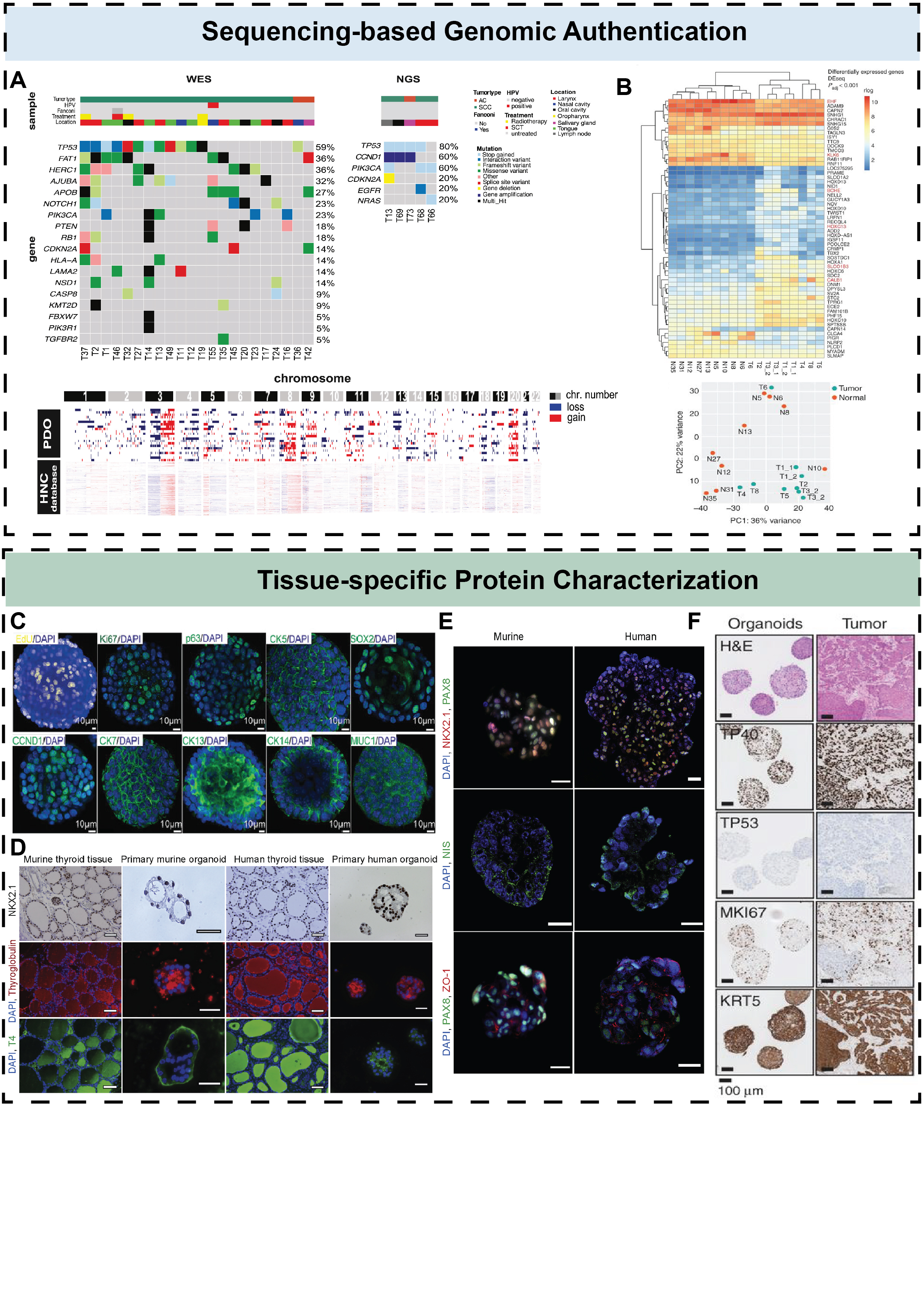
During organoid culture, cells aggregate and self-organize into defined three-dimensional structures. Microscopy often reveals morphological features that resemble those of the source tissue, enabling an initial assessment of fidelity by architecture. Investigators established a panel of eight PDO models that expanded efficiently in Matrigel (a basement-membrane extract enriched in laminin and type IV collagen) and typically formed dense solid or cystic spheroids with a peripheral basal-like layer and central differentiation[38]. Although organoid size inevitably shows some heterogeneity, it should be constrained within predefined limits, and overall morphology should be reproducible across batches. Architecturally, models should recapitulate native tissue features-stratification, apico-basal polarity, and luminal or follicle-like domains-which are key morphological readouts of maturation and functional fidelity. In practice, paired sections of the primary tissue and derived organoids can be processed and stained in parallel, with fluorescence labeling of tissue-specific proteins. Morphological and histopathological assessment offers low cost, rapid turnaround and broad applicability, which suits time-sensitive drug testing. After embedding and sectioning, routine H&E staining is used to review luminal or clustered architecture, epithelial polarity and cell–cell junctions, and to document keratinisation, gland formation, mucin secretion or colloid deposition[39]. Side-by-side comparison with the parental tumor is recommended, with semi-quantitative metrics such as lumen fraction, cellular density and polarity distribution. Nasopharyngeal organoids retain an inner layer of basal epithelial progenitors (p63+, CK5+) that also express stemness-associated markers SOX2 and CCND1. In parallel, they show multilineage differentiation, including CK7+ pseudostratified epithelium, CK13/CK14+ stratified squamous epithelium, and MUC1+ goblet cells. This lineage architecture indicates that the model reconstructs in vitro the basal-progenitor-to-differentiated hierarchy of the nasopharyngeal epithelium and can be used to study development and treatment responses[32] (Figure 3C). Immunolabeling of primary murine and human thyroid spheroids showed positive staining for NKX2.1 (also known as thyroid transcription factor-1), thyroglobulin, and T4, with calmodulin remaining negative, thereby confirming their thyroid origin (Figure 3D). Both murine and human thyroid organoids showed continuous ZO-1 tight-junction staining, indicating maintenance or restoration of thyroid epithelial barrier integrity (Figure 3E). A recent study showed that paraffin-embedded HNSCC organoids display, by immunohistochemistry, a peripheral layer of MKI67⁺/TP63⁺ basal cells and an inner compartment of KRT13⁺ differentiated cells, recapitulating the polarity and stratified architecture of the parent tissue and confirming successful organoid establishment with preservation of lineage features[35] (Figure 3F).
Organoid authentication uses two complementary tiers. Genomic profiling secures lineage identity and provenance, confirms driver retention, and measures concordance and subclones via STR or SNP fingerprinting plus WES or targeted panels with copy-number profiling, with single-cell or spatial assays as needed to monitor drift. Morphology and tissue-specific proteins give a rapid, low-cost readout of architecture and lineage using H&E and IHC/IF; in head and neck models this includes basal-to-differentiated layering in HNSCC, basal progenitors with multilineage programs in nasopharyngeal organoids, and thyroid lineage markers with intact tight junctions. Minimal quality control should predefine coverage and contamination thresholds, pass criteria, limited passages, and repeat genotyping before and after key assays. Table 2 compares genomic and histopathological modalities side-by-side, outlining scope, information content, sensitivity to heterogeneity, turnaround time, cost, scalability, typical readouts, and common pitfalls, and offers practical use-cases to guide method selection.
Table 2. Comparison of organoid characterization methods.
| Assay | Sample Input | Turnaround | Purpose | Cost |
|---|---|---|---|---|
| H&E staining | ++ | + | Compare tissue morphology | + |
| Immunohistochemistry (IHC) | ++ | + | Compare expression of tissue-specific proteins | + |
| Sanger sequencing | + | + | Detect tissue-specific genes (mutations) | + |
| Whole-exome sequencing (WES) | ++ | +++ | High-throughput profiling of characteristic gene variants and coverage | ++ |
| Transcriptome analysis (RNA-seq) | ++ | +++ | High-throughput assessment of gene expression and regulatory consistency | +++ |
| Whole-genome sequencing (WGS) | ++ | +++ | High-throughput genome-wide variant landscape and coverage | +++ |
| DNA methylation analysis | + | ++ | Epigenetic profiling and comparative analysis | ++ |
| STR profiling (authentication) | +++ | + | Verify sample origin and passage-to-passage genetic stability | + |
Note: "+ / ++ / +++" indicate relative requirement or intensity—more "+" means a larger sample input, a longer turnaround time, and a higher cost.
Applications of tumor organoids in head and neck diseases
Basic tumor biologyOncogenesis, tumor progression and metastasis form a continuous, interrelated process. During tumor initiation, organoid technology can recapitulate stem-cell behaviour, permit interrogation of signalling pathways and assess the effects of gene regulation on initiation and progression, thereby elucidating disease mechanisms. Organoid cultures retain, to a remarkable extent, the in vivo biological properties and genetic features of tumor cells, addressing limitations of conventional tumor models[40]. Everaging organoid technology, Fujii et al.[41]reconstructed the stepwise trajectory of colorectal cancer from initiation to progression. In gastric cancer, investigators established a large PDOs biobank and subsequently identified and validated genomic and molecular features predictive of chemotherapy response; co-culture with cancer-associated fibroblasts (CAFs) further underscored the critical role of the tumor microenvironment in mediating drug resistance. Together, these findings indicate that PDO models provide robust platforms for anticancer drug screening and response prediction[42]. By establishing a mouse oesophageal squamous cell carcinoma (ESCC) organoid model and integrating lineage-tracing techniques, Whelan K.A. et al.[43] demonstrated that ESCC cells originate from basal keratinocytes. At the metabolic level, oral squamous cell carcinoma (OSCC) organoids indicate that lactate promotes stem-like phenotypes, nominating the monocarboxylate transporter MCT1 as a potential target[44]. HPV-negative HNSCC PDOs have been established that faithfully recapitulate three-dimensional invasion in vitro. This platform enables parallel comparison of distinct invasion modes under a uniform genetic background and can be used for mechanism dissection, prognostic stratification, and screening of candidate therapies focused on YAP and matrix mechanics[38]. Researchers established a broad panel of patient-derived endocrine organoids from benign and malignant tumors of the thyroid, parathyroid, and adrenal glands, and—using functionally intact parathyroid PDOs as a model-combined mass-spectrometry-based metabolomics with live-cell flux analyses to show their utility for metabolic and bioenergetic screening in primary hyperparathyroidism (PHPT), with organoid metabolic profiles recapitulating those of matched patient tissues[45]. Using patient-derived TC organoids, investigators modeled chronic low-dose exposure to perfluoroalkyl carboxylic acids and quantified molecular responses by Western blotting, immunofluorescence, and high-content imaging. Long-chain PFACs altered thyroid lineage markers, epithelial–mesenchymal transition programs, and Ki-67 proliferation within organoids, demonstrating that organoids provide a practical system to assess carcinogenic effects of environmental exposures and to screen interventions that might reduce them[46]. Furthermore, Studies have shown that 3D SCC organoids derived from cell lines, PDXs, and patient biopsies reveal ethanol-induced mitochondrial oxidative stress that kills bulk tumor cells, while CD44+ high stem-like cells persist via autophagy. Blocking autophagy in these organoids restores ethanol sensitivity and curbs xenograft growth, underscoring organoids as a practical platform to dissect alcohol-linked biology and test autophagy-targeted therapies[47].
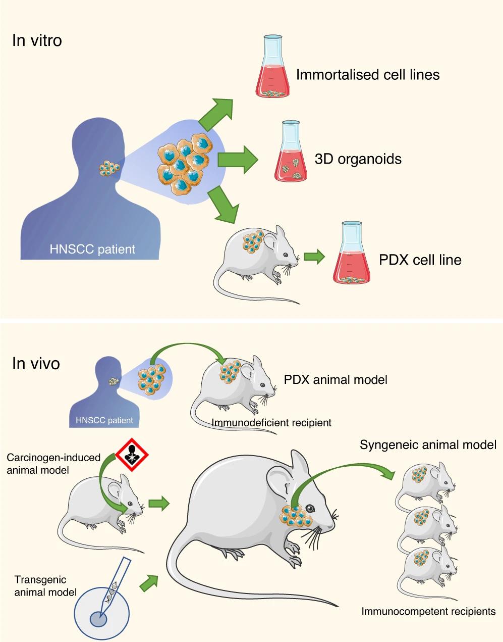
In microenvironment and angiogenic control, tri-culture organoid systems containing endothelial cells, cancer-associated fibroblasts and cancer cells identify stromal nicotinamide N-methyltransferase (NNMT) as a stabiliser of angiogenesis, revealing a stromal target for anti-angiogenic therapy[48]. In pathway remodelling, loss of SMAD4 or inhibition of TGFβ signalling enhances WNT activity and remodels the extracellular matrix in organoids; murine oral tumor organoids thereby acquire genomic and histological features that closely mirror human HNSCC, supporting a tumor-suppressive role for SMAD4[49] (Figure 4). Using HNSCC PDOs alongside cell lines and xenografts, PRMT1 was shown to confer carboplatin resistance by recruiting SWI/SNF via SMARCC1 and activating IGF2BP2. Genetic loss or organoid-level disruption of the PRMT1–SMARCC1 interaction suppressed growth and restored carboplatin sensitivity, revealing a vulnerability independent of PRMT1 catalytic activity that is amenable to protein–protein interaction inhibitors or targeted degraders[50].
Figure 4. Above, in vitro preclinical models include immortalised cell lines, 3D organoids and PDX cell lines. Below, in vivo murine preclinical models include PDX, syngeneic, carcinogen induced and transgenic animal models.Copyright © 2023, Patricia Chaves.
Head and neck tumor organoids provide an experimentally tractable system to interrogate tumor biology: they reproduce metabolic adaptation, stemness and epithelial-to-mesenchymal transition (EMT), stromal and vascular crosstalk, pathway rewiring, radiosensitivity and chromatin-mediated drug resistance in a patient-specific manner, allowing direct causal testing of these processes in vitro. By linking molecular perturbations to functional phenotypes within a clinically faithful model, they have become a central platform for mechanistic dissection of head and neck cancer.
Drug screening and new drug developmentCompared with traditional two-dimensional cell lines, three-dimensional tumor organoids models have lower sensitivity to drugs and are closer to the actual situation in clinical practice[51, 52] . In a population with pronounced interpatient variability, PDOs provide a quantitative and verifiable route to shortlist monotherapies, combinations and radiotherapy schedules. For early response assessment, Shah and colleagues combined optical metabolic imaging with PDOs and detected treatment effects after one day of exposure, with proliferation, apoptosis and xenograft volume used as parallel gold standards, which suits high-throughput prescreening and regimen convergence[53]. For cytotoxic agents, the CTOS-PDOs platform from Tanaka’s group compared cisplatin and docetaxel[17], and in vitro IC50 values aligned with in vivo response direction, underscoring substantial interpatient heterogeneity and enabling selection of chemotherapy backbones. For targeted and novel strategies, Driehuis and colleagues used HNSCC PDOs to test EGFR-targeted photodynamic therapy with antibody- or nanobody-photosensitiser conjugates, observing efficacy proportional to EGFR expression[51]; PDOs from normal oral mucosa expressed less EGFR, supporting biomarker-based stratification and more selective payload delivery to widen the therapeutic window.
When tissue is scarce in advanced disease, CTC-derived PDO enable micro-sampling with rapid feedback; in a head and neck cancer cohort a multivariable model based on CTC-PDO responses predicted platinum sensitivity with 93.75% accuracy, indicating real-world utility[34]. Pauli et al. have used several cancer types from different anatomical locations to establish tumor organoids[54]. WES was performed to confirm genetic similarities between tumor organoids and primary tumors (96%). In addition, there is a push to use known gene–drug associations (n = 160) in tumor organoids using high-throughput drug screening with genomic analysis[54]. Similarly, some studies have predicted outcomes of drug treatments using genomic analyses of the cancer organoids[55]. In addition to established treatments, several drugs have recently been developed and are undergoing trials for HNC. These new treatments are currently in Phase 1 and Phase 2 clinical trials with most focus on the development of targeted therapeutic agents, which can be used in combination with conventional therapies. Some of these targeted therapies that are in clinical trials include erlotinib, ABT-510 and bevacizumab, which are novel therapies for HNC.
Based on the clinical trial website (https://clinicaltrials.gov) [56], there are 2670 clinical studies recorded (to test current treatment combinations as well as novel treatments), while only 1085 were completed globally. Among the completed studies, 248 have shown favourable outcomes including improved overall survival (e.g.: Pemetrexed plus Gemcitabine), lower rate of recurrence (e.g., synergistic effect of Cetuximab, Hydroxyurea, Fluorouracil and radiotherapy), the low incidence rate of non-haematologic and haematologic toxicity side effects (e.g., synergistic effect of Kanglaite and chemotherapy) etc.
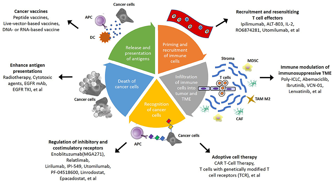
Immune-related studiesThe tumor microenvironment, including its immune compartment, shapes initiation, progression, treatment response and resistance in HNC[57, 58] Figure 5. Conventional HNSCC organoids are largely epithelial and do not capture immunosuppressive ecology. Two complementary platforms are therefore used. Organoid co-culture can serve as a pre-stratification tool to identify likely beneficiaries and to optimise combinations. However, significant challenges remain in modeling the immune microenvironment. A major limitation is the lack of a functional vascular system, which restricts the infiltration of immune cells into the organoid core. Furthermore, in co-culture systems using non-autologous immune cells, human leukocyte antigen mismatching can trigger allogenic responses that confound true tumor-specific cytotoxicity. Finally, maintaining the long-term viability of immune cells (particularly T cells) in organoid media is difficult, as conditions optimized for epithelial growth may not support sustained immune activation, leading to premature T-cell exhaustion. Future iterations utilizing microfluidic platforms and vascularized chips are needed to overcome these barriers. In reconstructed co-cultures, PDOs embedded in matrix are cultured with autologous peripheral or tumor-infiltrating immune cells to quantify cytotoxicity, cytokine release, infiltration depth and exhaustion phenotypes. Using this approach, Dijkstra and colleagues generated tumor-reactive T cells that selectively killed matched organoids, supporting prescreening of PD-1-based strategies[59]. In native microenvironment–preserving systems, air–liquid interface culture and microfluidic channel models retain endogenous T, B, NK together with stromal elements[60]. l-Samadi et al. manufactured the frst in vitro fully humanized 3D microfuidic chip using patient-derived cancer cells, patients’serum, and immune cells to test personalized immunotherapeutics for HNSCC patients[61]. Neal and colleagues, in human and murine tumors, showed expansion and activation of antigen-specific lymphocytes under PD-1 or PD-L1 inhibition with restoration of cytotoxic activity[62]. These frameworks also permit testing of microenvironmental drivers. In a tongue cancer organoid system, Sawant and colleagues showed that cancer-associated fibroblasts promote stage-dependent epithelial malignant transition, revealing intervention points across the stroma–immune–tumor axis[63]. Clinically, although PD-1/PD-L1 inhibitors improve long-term survival in a subset, durable responses remain uncommon due to defects in antigen presentation and activation of suppressive pathways[64]. Organoid co-culture can serve as a pre-stratification tool to identify likely beneficiaries and to optimise combinations and dosing schedules[65]. However, significant challenges remain in modeling the immune microenvironment[62]. A major limitation is the lack of a functional vascular system, which restricts the infiltration of immune cells into the organoid core[66]. Furthermore, in co-culture systems using non-autologous immune cells, HLA mismatching can trigger allogenic responses that confound true tumor-specific cytotoxicity[65]. Finally, maintaining the long-term viability of immune cells (particularly T cells) in organoid media is difficult, as conditions optimized for epithelial growth may not support sustained immune activation, leading to premature T-cell exhaustion[65]. Future iterations utilizing microfluidic platforms and vascularized chips are needed to overcome these barriers[67].
Figure 5. Schematic summary of potential strategies to overcome immunosuppressive TME in HNSCC. Copyright © 2019, Wang Hui-Ching.
HNC is strongly shaped by population differences, HPV status and lifestyle exposures, with pronounced heterogeneity at phenotypic and genomic levels. Building systematic, high-fidelity biobanks of PDOs is therefore essential.
PDOs combine the utility of personalised drug testing with mechanistic interrogation of key drivers and thus form the core of large head and neck cancer repositories. Such biobanks support comparative assessment of drug responsiveness, functional evaluation of new agents, harmonisation of regimen suitability and mechanistic studies of tumor initiation and progression [11, 35]. Owing to disease biology, technical complexity and resource demands, reported head and neck organoid biobanks remain modest in size, yet their translational value is increasing.
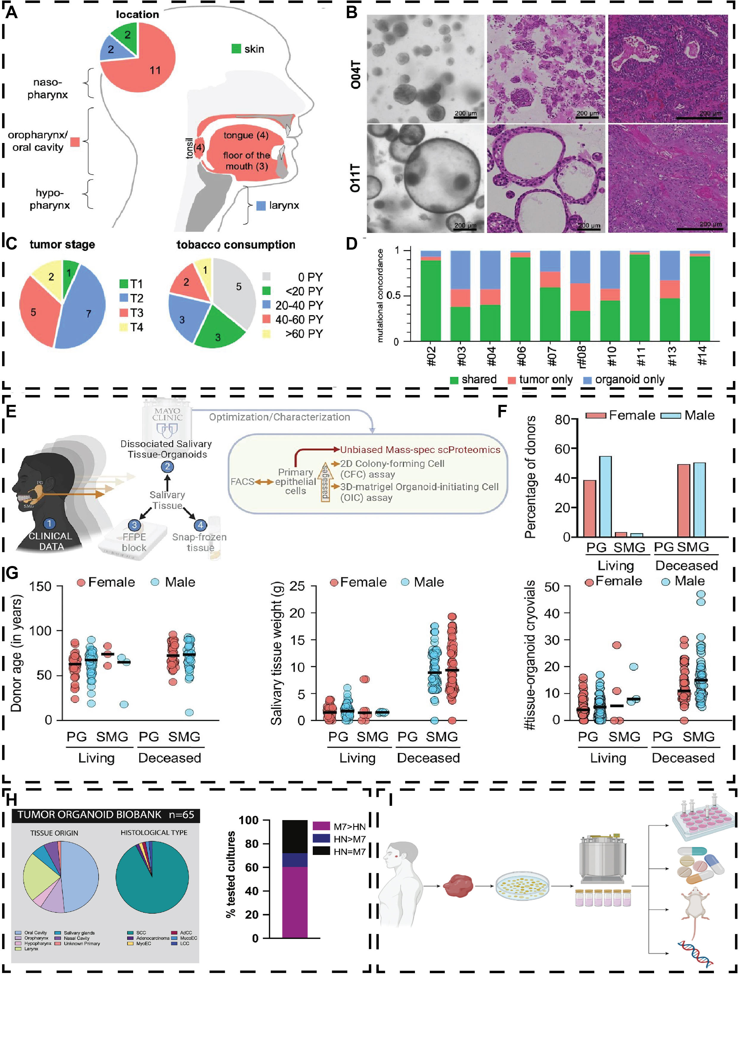
Under unified workflows, biobanks can run drug and radiosensitivity profiling, functional genetic screens and multi-omics, and align in vitro readouts with clinical outcomes for biomarker discovery and stratified validation. For example, a head and neck organoid biobank (HNOB) capturing TP53-mutant and HPV16-driven subtypes has been used for patient stratification and for mechanistic validation of driver-specific treatment responses[68] (Figure 6A-D). An organoid repository spanning benign and malignant salivary tumors recapitulated the transcriptomic and histological features of primaries and nominated PTP4A1 as a candidate diagnostic marker for mucoepidermoid carcinoma[69]. A diverse, clinically annotated salivary regenerative biobank established at the Mayo Clinic provides a platform for studying salivary gland stem and progenitor cells and for developing regenerative therapies[70](Figure 6E-F).
Figure 6. Clinical and genetic features of the HNC organoid biobank. (A) Tumor sites. (B) Cohort clinical parameters. (C) Representative mor-phology and H&E of compact (O04T) and cystic (O11T) organoids with matched tumor histology; scale bars. (D) WES-based mutational concor-dance between organoids and paired tumors for SNPs with VAF > 0.05. Copyright © 2025, Issing, C. (E) Workflow for processing parotid (PG) and submandibular (SMG) tissues to derive organoids, followed by cryopreservation and biobanking from living and deceased donors of both sexes. (F) H&E of PG and SMG tissues and matched organoids showing ductal and acinar architecture. (G) Donor age distribution and tissue weights at receipt for PG and SMG, stratified by donor status and sex.Copyright © 2024, Aalam.
Key bottlenecks persist. Small specimen size, abundant stroma and marked cellular heterogeneity complicate model establishment; insufficient tumor cellularity is a major cause of failure. When the variant allele fraction of driver mutations increases from less than 0.15 to more than 0.3, establishment success rates rise from about 28% to about 77%[71]. Additional constraints include complex procurement and processing workflows, limited specialised teams and shared infrastructure, and the absence of centralised databases, harmonised standards and quality control[72]. Institutions often differ in culture methods, data capture and QC metrics. We recommend standard operating procedures and QC frameworks that cover procurement, media composition, passage limits, sequencing depth and coverage, control sets and pass criteria, together with central databases and external quality assessment. Integration of clinical annotation with multi-omics will increase comparability and portability and will position organoid biobanks as core platforms for pre-treatment stratification and prospective biomarker validation in head and neck cancer.
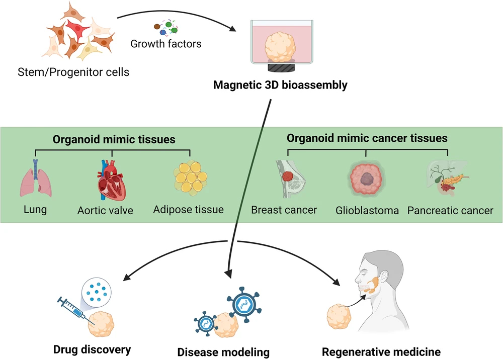
Regenerative medicineOrganoid technology promises to transform biomedical research by providing physiologically relevant models for human development, disease mechanisms and therapeutic discovery. Derived from stem cells, organoids self-organise into three-dimensional tissues that recapitulate the architecture and functions of their in vivo counterparts. Radiotherapy for head and neck malignancies damages the salivary glands and commonly causes xerostomia[73]. Bücheler et al. cultured salivary gland organoids on microcarriers, introducing a concept for functional reconstruction[74]. Ozdemir et al. generated salivary microtissues from primary human salivary gland myoepithelial cells (hSMECs) and stem/progenitor cells isolated from normal tissue; across serial passages, the myoepithelial phenotype remained stable and resembled that of other exocrine epithelia[75]. Furthermore, magnetic 3D bioassembly (M3DB) and microfluidic coaxial 3D bioprinting, under matrix-free conditions, drive adult stem cells and primary salivary gland cells to self-organise into functional micro-organoids with luminal and ductal architectures; these constructs exhibit secretory responses to cholinergic and adrenergic stimulation, and organoid-derived extracellular vesicles promote in vitro repair of irradiated salivary glands[76](Figure 7).
Figure 7. M3DB bioprinting produces tissue-mimetic organoids-lung, aortic valve, adipose, and cancers (breast, pancreatic, glioblasto-ma)-for drug discovery, disease modeling, and regenerative medicine.Copyright © 2024, Klangprapan.
Summary and Perspectives
PDOs of head and neck cancer are emerging as versatile three-dimensional models that address key limitations of conventional two-dimensional cell lines in architecture, genetic heterogeneity and clinical reproducibility. Their capacity for long-term propagation, cryopreservation and recovery supports the creation of organoid biobanks that enable genomic and transcriptomic analyses, mutation discovery and large-scale drug sensitivity and resistance testing. On the translational front, salivary-gland organoids offer a route to functional restoration after radiotherapy, with the potential to alleviate xerostomia. Efforts to recapitulate the tumor microenvironment have advanced through co-culture and air–liquid interface systems that incorporate cancer-associated fibroblasts and stromal counterparts. Integration with microfluidic platforms, organ-on-chip devices and tissue assemblies further permits controlled simulation of perfusion, chemical gradients and mechanical cues, thereby improving the external validity of treatment responses.
A central tension remains between model fidelity and scalability. As complexity rises, so do costs, technical barriers and batch-to-batch variability, which hinder high-throughput use and rapid preclinical decision-making. A pragmatic near-term priority is to establish standardized pipelines with rigorous quality control (QC). To improve reproducibility across centers, we propose that future studies must report a minimum set of parameters: (1) specific establishment rate and doubling time; (2) precise growth factor concentrations and matrix stiffness; (3) passage number used for experiments (ideally <10 to minimize drift); and (4) concordance metrics (e.g., STR profiling or SNP array) comparing the organoid to the original tissue. These standards will facilitate the creation of interoperable, multi-site biobanks.
Looking ahead, progress is likely to hinge in three directions. First, systematic and tunable incorporation of microenvironmental elements: beyond existing co-culture and air–liquid interface approaches, adding macrophages, lymphocytes, endothelial cells and neurons on perfusable microfluidic platforms will allow coordinated modelling of immune, vascular and neural axes. Second, standardisation at scale: cross-centre standard operating procedures, unified multi-omics and pharmacology readouts and strategies to minimise batch effects will enable interoperable, multi-site biobanks. Third, clinical integration: to deliver personalised care, workflows must shorten sample-to-result turnaround and be tested prospectively, with endpoints that link PDO-guided therapy selection to patient outcomes. Coordinated evaluation with radiotherapy, immunotherapy and targeted agents will be essential.
With these elements in place, HNC PDOs should better expose mechanisms of initiation, progression and metastasis, underpin high-throughput drug discovery and combination testing, and, in the clinic, support the selection and verification of personalised treatment strategies, advancing precision oncology.
Abbreviations
ABT - Antibiotic Therapy; BDJ - British Dental Journal (journal); BRAF - B-Raf proto-oncogene serine/threonine kinase; CNV - Copy Number Variation; CTC - Circulating Tumor Cell; CTC-PD-TO - Circulating Tumor Cell-derived Patient-Derived Tumor Organoid; CTOS - Cancer Tissue-Originated Spheroids; DNA - Deoxyribonucleic Acid; DOI - Digital Object Identifier; EBER - Ep-stein–Barr virus-encoded small RNA; EBV - Epstein–Barr Virus; EGF - Epidermal Growth Factor; EGFR - Epidermal Growth Fac-tor Receptor; EMT - Epithelial–Mesenchymal Transition; ESCC - Esophageal Squamous Cell Carcinoma; FGF - Fibroblast Growth Factor; GEM - Gemcitabine (chemotherapeutic drug); HNC - Head and Neck Cancer; HNOB - Head and Neck Organ-oid Biobank; HNSCC - Head and Neck Squamous Cell Carcino-ma; HPV - Human Papillomavirus; IF - Immunofluorescence; IHC - Immunohistochemistry; IV - Intravenous; NGS - Next-Gen-eration Sequencing; NIS - Sodium/Iodide Symporter; NK - Nat-ural Killer (cell); NNMT - Nicotinamide N-methyltransferase; NPC - Nasopharyngeal Carcinoma; NTRK - Neurotrophic Tyro-sine Receptor Kinase; OSCC - Oral Squamous Cell Carcinoma; PCA - Principal Component Analysis; PD - Progressive Disease; PDO - Patient-Derived Organoid; PDX - Patient-Derived Xeno-graft; PDXO - Patient-Derived Xenograft Organoid; PG - Parotid Gland; PHPT - Primary Hyperparathyroidism; QC - Quality Con-trol; RET - RET proto-oncogene (rearranged during transfec-tion); RNA - Ribonucleic Acid; SCC - Squamous Cell Carcinoma; SGC - Salivary Gland Carcinoma; SLAS - Society for Laboratory Automation and Screening; SMG - Submandibular Gland; SNF - Sucrose Non-Fermentable (part of SWI/SNF chromatin re-modeling complex); SNP - Single Nucleotide Polymorphism; STAR - Spliced Transcripts Alignment to a Reference (RNA sequencing aligner); STR - Short Tandem Repeat; SWI - Switch (part of SWI/SNF chromatin remodeling complex); TC - Thyroid Carcinoma; TGF - Transforming Growth Factor; TGM - Transglu-taminase; TME - Tumor Microenvironment; VAF - Variant Allele Frequency; WES - Whole Exome Sequencing; WNT - Wingless/Integrated (Wnt signaling pathway); WR - Ward Round; YAP - Yes-Associated Protein; ZO - Zonula Occludens (tight junction protein)
Declarations
Author Contributions
Fangzheng Yi: Conceptualization, Investigation, Writing - original draft, Writing - review & editing.
Acknowledgements
Not Applicable.
Funding information
Not Applicable.
Ethics Approval and Consent to Participate
Not Applicable.
Competing Interests
The authors declare that they have no existing or potential commercial or financial relationships that could create a conflict of interest at the time of conducting this study.
Data availability
All data discussed in this review are derived from previously published studies and are available in the cited references. No new data was generated or analysed in this study.
References
 Figures
 References
 Peer
Peer Information
Figure 1. Schematic of the sources and applications of tumour organ-oids in head and neck diseases (Created with bioRender.com).
Figure 2. (A) Establishment of organoids from HNC tissue, and HNC organoids recapitulate thecharacteristics of original tumor tissues. Copyright © 2018 Noriaki Tanaka. (B) Schematic representation of murine and human thyroid primary cell culture. Thyroid gland tissue was mechanically and enzymatically digested and resuspended in culture medium or seeded in Matrigel. Copyright © 2021 Vivian M.L. (C) Mouse thyroid cells (n=3) after mechanical-enzymatic dissociation cultured in TGM; spontaneous spheroids at 24 h. Representative bright-field and 72 h high-magnifica-tion images. (D) Workflow for constructing primary and orthotopic NPC models, time-course bright-field images showing a monoclonal organoid arising from a single cell and representative H&E image of normal nasopharyngeal organoids. Copyright © 2024 Xudong Wan. (E) Experimental schematic for murine salivary gland organoids: parotid (mPG), sublingual (mSLG), and submandibular (mSMG) amd mSMG organoids cultured in complete growth expansion medium (GEM) or GEM lacking individual factors, with growth monitored by brightfield microscopy. Copyright © 2022, Yoon.
Figure 3.(A) Genomic features of HNC organoids. DNA sequencing of 35 organoid cultures (WES, n=30; targeted hotspot panel, n=5) is sum-marized as a mutation oncoplot, with high-level copy-number variation (CNV) profiles shown and compared against reference datasets from pri-mary HNC tumors. Red indicates chromosomal amplifications, and blue indicates deletions. Copyright © 2023 Millen Rosemary. (B) A heat map displays 58 differentially expressed genes between groups (Padj < 0.001; DESeq2); blue indicates low expression, red indicates high. RNA-seq separates tumor and normal organoids. PCA of RNA-seq profiles from normal wild-type organoids (n=9, orange) and tumor-derived organoids (n=7, blue) shows clear group separation. Copyright © 2019 Driehuis Else. (C) Confocal IF of organoids showing EdU incorporation and markers: Ki67 (proliferation), p63/CK5 (basal), SOX2/CCND1 (stemness), CK7 (pseudostratified epithelium), CK13/CK14 (stratified squamous), and MUC1 (goblet). Copyright © 2024 Wan Xudong. (D) NKX2.1, thyroglobulin, and T4 staining of murine and human tissue and primary murine spheres after 1 day in floating culture and primary human thyroid organoids after 7 days in culture in Matrigel shows a nuclear staining for NKX2.1 and staining for thyroglobulin and T4.© 2021 Vivian M.L. (E) Representative confocal images of immunofluorescence staining for NKX2.1, PAX8, NIS, and ZO-1 in murine and human organoids. Copyright © 2021 Vivian M.L. (F) Hematoxylin and eosin (H&E) staining and immunostaining for basal cell marker TP40, tumor suppressor TP53, proliferation marker MKI67 and KRT5 of paraffin-embedded T15 organoids and corresponding tissue. Copyright © 2019 Driehuis.
Figure 4. Above, in vitro preclinical models include immortalised cell lines, 3D organoids and PDX cell lines. Below, in vivo murine preclinical models include PDX, syngeneic, carcinogen induced and transgenic animal models.Copyright © 2023, Patricia Chaves.
Figure 5. Schematic summary of potential strategies to overcome immunosuppressive TME in HNSCC. Copyright © 2019, Wang Hui-Ching.
Figure 6. Clinical and genetic features of the HNC organoid biobank. (A) Tumor sites. (B) Cohort clinical parameters. (C) Representative mor-phology and H&E of compact (O04T) and cystic (O11T) organoids with matched tumor histology; scale bars. (D) WES-based mutational concor-dance between organoids and paired tumors for SNPs with VAF > 0.05. Copyright © 2025, Issing, C. (E) Workflow for processing parotid (PG) and submandibular (SMG) tissues to derive organoids, followed by cryopreservation and biobanking from living and deceased donors of both sexes. (F) H&E of PG and SMG tissues and matched organoids showing ductal and acinar architecture. (G) Donor age distribution and tissue weights at receipt for PG and SMG, stratified by donor status and sex.Copyright © 2024, Aalam.
Figure 7. M3DB bioprinting produces tissue-mimetic organoids-lung, aortic valve, adipose, and cancers (breast, pancreatic, glioblasto-ma)-for drug discovery, disease modeling, and regenerative medicine.Copyright © 2024, Klangprapan.
Peer-review Terminology
Identity transparency: Single anonymized
Reviewer interacts with: Editor
Details
This is an open access article under the terms of the Creative Commons Attribution License(http://creativecommons.org/licenses/by/4.0/), which permits use, distribution and reproduction in any medium, provided the original work is properly cited.
Publication History
Received 2025-11-12
Accepted 2025-11-23
Published 2025-12-14