




Abstract
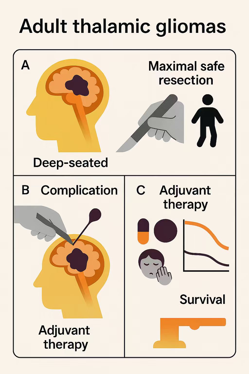
Objective: This systematic review and meta-analysis evaluated surgical outcomes, complications, and survival in adult thalamic glioma patients to inform optimal surgical and adjuvant treatment strategies.
Methods: We searched PubMed, Web of Science, and Embase up to July 10, 2025, analyzing 15 studies with 695 adults. Data included patient characteristics, surgical approaches (lateral, medial, posterior), extent of resection (EOR, gross total/subtotal resection [GTR/STR] or partial resection [PR]), complications, and overall survival (OS). Cox proportional hazards models assessed survival factors (p<0.05).
Results: High-grade gliomas (HGGs) accounted for the majority (77.8%), and GTR/STR was achieved in 86.1% of cases, primarily through a lateral-related surgical approach (51.8%). Common postoperative complications were movement disorders (29%), infection/fever (25%), and sensory disorders (13%). In the pooled cohort consisting of 103 patients, median OS was 16 months for HGGs and 23 months for low-grade gliomas (LGGs). Multivariate Cox analysis confirmed prolonged OS in LGGs (hazard ratios [HRs]: 0.34, p = 0.005) and with adjuvant therapies (chemotherapy: HR 0.19, p=0.025; radiotherapy: HR 0.10, p=0.002; radiochemotherapy: HR 0.13, p=0.003).
Conclusion: Maximal safe resection with adjuvant therapies enhances survival in thalamic gliomas, despite high complication rates. Tailored surgical strategies and molecular profiling are essential to optimize outcomes and guide targeted therapies.
Keywords: Thalamic gliomas, Surgical resection, Postoperative complications, Adjuvant therapy, Overall survival
Introduction
Thalamic gliomas are rare brain tumors originating in the thalamus, a critical midline structure within the diencephalon, situated bilaterally adjacent to the third ventricle. The thalamus is anatomically delineated as follows: anteriorly by the anterior tubercle, posteriorly by the pulvinar, medially by the third ventricle, and laterally by the internal capsule, septum, and basal ganglia. Superiorly, it is bordered by the lateral ventricles, while inferiorly, it abuts the hypothalamus and midbrain. As a pivotal deep brain structure, the thalamus serves as a relay hub, facilitating the transmission of sensory and motor signals[1]. Thalamic gliomas constitute approximately 1–5% of all intracranial neoplasms in adults, posing significant diagnostic and therapeutic challenges due to their deep-seated location and close proximity to critical neural pathways[2]. In the 5th edition of the World Health Organization (WHO) central nervous system (CNS) classification, H3K27M mutations are recognized as a defining diagnostic marker for diffuse midline gliomas (DMG). These mutations are detected in 78–90% of thalamic and brainstem gliomas and are strongly correlated with an unfavorable clinical prognosis, with a median overall survival of less than 12 months[3-5]. The intricate anatomy of the thalamus poses substantial challenges for surgical management of thalamic lesions. Despite these difficulties, surgery remains the cornerstone of treatment, with the primary goal of achieving maximal safe resection while preserving neurological function[6]. The deep-seated location of these lesions heightens the risk of postoperative complications, including hemiparesis, sensory deficits, and cognitive impairments, which can profoundly impact patients' quality of life[7].
The rarity of thalamic gliomas has constrained the development of large-scale studies, with current evidence primarily drawn from small case series or retrospective analyses[8, 9]. Surgical resection remains the cornerstone of treatment, yet its efficacy and safety are debated due to variability in outcomes and the potential for significant morbidity[10]. Although advances in neuroimaging, stereotactic techniques, and intraoperative monitoring have improved surgical precision, the differences in surgical approaches, ranging from aggressive GTR to more conservative STR and PR, persist across institutions[11, 12]. These disparities highlight the urgent need for a comprehensive evaluation of surgical outcomes and associated complications.
The heterogeneity in tumor histology, patient demographics, and adjuvant therapies, such as radiotherapy and chemotherapy, complicates the interpretation of existing data[10, 13-15]. To address these challenges, we conducted a systematic review and meta-analysis to comprehensively evaluate surgical outcomes and complications in adult patients with thalamic gliomas. By synthesizing data from diverse studies, we aimed to assess the surgical outcomes and the role of adjuvant therapies, with the ultimate goal of informing clinical decision-making and enhancing patient care.
Materials and Methods
This systematic review and meta-analysis was conducted according to the Preferred Reporting Items for Systematic Reviews and Meta-Analyses (PRISMA) guidelines[16]. The quality of included studies was evaluated according to design characteristics (a retrospective cohort, consecutive enrollment, and defined outcome criteria).
Literature Retrieval and Search Strategies.
We conducted a comprehensive literature search across PubMed, Web of Science, and Embase up to July 10, 2025, to identify studies relevant to the evaluation, synthesis, and analysis of surgical techniques and associated complications in adult thalamic gliomas. The search strategy was meticulously designed, integrating a combination of Medical Subject Headings (MeSH), Emtree terms, and additional standardized keywords, with Boolean operators strategically employed to optimize the combination of search terms. The primary search criteria centered on the MeSH terms "thalamic glioma" and "surgical treatment." Detailed search terms and Boolean strings used for each database are available upon request to ensure reproducibility. Additionally, we performed a thorough citation analysis of all retrieved articles to identify further relevant studies, ensuring a robust and inclusive review of the literature.
Inclusion and Exclusion Criteria.
Studies were included in the review if they fulfilled the following criteria: (1) the study cohort included a minimum of three patients, (2) the cohort consisted of adult patients diagnosed with thalamic glioma who underwent surgical intervention, and (3) postoperative clinical outcome data for the cohort were reported. Studies were excluded if they met any of the following criteria: (1) the publication was a case report, editorial, conference abstract, non-English language study, or involved animal subjects, or (2) the study presented unclear or ambiguous data.
Data Extraction Procedure.
Data extraction and review were independently conducted by two investigators (H. Mao and F. Mu). In instances of disagreement, a third investigator (M. Yang) provided independent adjudication to achieve consensus. Extracted data included, but were not limited to, the following variables: cohort size, patient age, sex, preoperative symptoms, tumor laterality, surgical approach, histological findings, WHO grade, EOR, postoperative adjuvant therapies, postoperative complications, and OS.
To address inconsistencies in terminology across studies, surgical approaches were categorized into three standardized groups: lateral, medial, and posterior approaches. Similarly, due to variability in the definition of EOR, GTR and STR were combined into a single category for statistical analysis. For the WHO classification of gliomas, grades 1 and 2 were designated as LGGs, while grades 3 and 4 were classified as HGGs. Individual patient data from the included studies were systematically compiled and summarized to facilitate further analysis.
Statistical Analysis.
Continuous variables were presented as means ± standard deviations (SD), while categorical variables were presented as frequencies (n). In the presence of substantial heterogeneity (I² > 50%), a random-effects model was employed to estimate the pooled postoperative complication rate using RevMan software (version 5.4). Individual patient data were aggregated and analyzed using Cox proportional hazards models for both univariate and multivariate analyses, performed in the open-source statistical software R (version 4.1.2). Statistical significance was established using a two-tailed p-value threshold of < 0.05 for all tests.
Results
Literature review results.
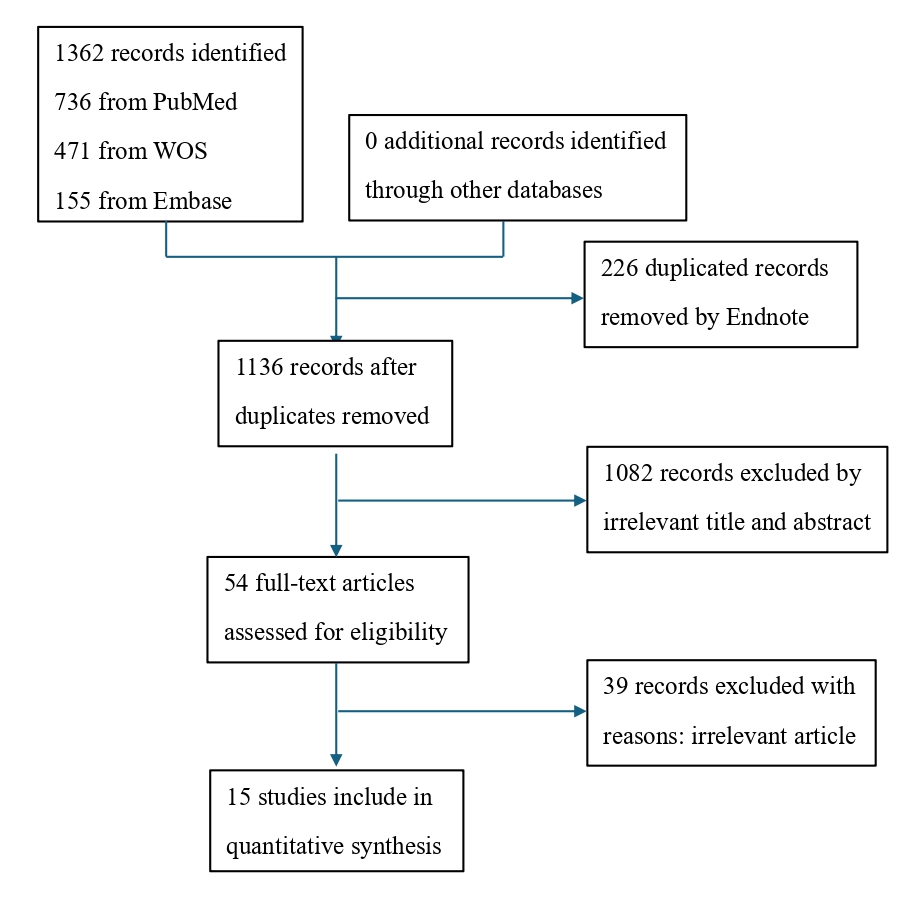
Using a predefined search strategy, a total of 1,362 articles were retrieved, including 736 from PubMed, 471 from Web of Science, and 155 from Embase. Duplicate records (n=226) were removed using EndNote software. Following title and abstract screening, 1,082 articles were excluded due to their lack of relevance to the research topic. The remaining 54 articles underwent full-text review, with 39 deemed irrelevant. Ultimately, 15 articles were included for quantitative analysis. The complete search and screening process is depicted in Figure 1.
Figure 1. Flowchart of studies that could be quantitatively synthesized according to the inclusion and exclusion criteria.
Clinical characteristics of patients.
A total of 15 studies were included in this analysis. Pediatric patients were excluded, resulting in a cohort of 695 adult patients. HGG predominated in histological grading, comprising 541 cases (77.8%), with four studies exclusively reporting HGG cases[9, 15, 17, 18]. On this basis, studies reported H3K27M mutation (n=67, 20.6%)[9, 19-22], IDH1/2 mutation (n=19, 5.8%)[9, 19-22], TP53 mutation (n=160, 49.2%)[9, 19-22], and MGMT methylation (n=135, 43.4%)[9, 15, 19, 21, 22]. Tumor laterality was reported in 461 cases, with 212 (46.0%) located in the left thalamus, 242 (52.5%) in the right thalamus, and 7 (1.5%) involving both thalami. A summary of each study is presented in Table 1. Additionally, six studies provided detailed individual patient data[9, 11, 15, 18, 23, 24], these data will be utilized for subsequent Cox proportional hazards analysis.
Table 1. Summary of 15 studies of thalamic gliomas.
| Study | No. of Patients | Age, mean±sd | Gender, M/F | Histology, n | WHO Grade, n | Side of tumor, n | Intrathalamic Location, n |
|---|---|---|---|---|---|---|---|
| Kis et al., 2014 | 5 | 34.2 ± 17.6 | 4/1 | FA: 1 GBM: 3 PA: 1 |
LGG: 2 HGG: 3 |
Left: 2 Right: 3 |
Medial thalamus: 2 Posterior thalamus: 3 |
| Steiger et al., 2000 | 9 | 40.6 ± 13.8 | NR | AA: 1 G: 3 PA: 4 |
LGG: 5 HGG: 4 |
Left: 5 Right: 4 |
NR |
| Majchrz et al., 2018 | 36 | 37, NR | 20/16 | AA: 16 AOA: 1 BM: 1 FA: 4 GBM: 1 OA: 4 PCA: 7 PMA: 1 |
LGG: 13 HGG: 23 |
Left: 6 Right: 30 |
NR |
| Niu et al., 2020 | 102 | 40 ± 14.4 | 56/46 | GBM: 62 Other types: 40 |
LGG: 14 HGG: 88 |
Left: 57 Right: 41 Both: 4 |
Confined in thalamus: 40 Beyond thalamus: 62 |
| Cao et al., 2015 | 111 | 33.4 ± 13.2 | 71/40 | A: 88 GG: 2 OA: 17 |
LGG: 65 HGG: 49 |
Left: 51 Right: 60 |
Anterior: 20 Lateral: 7 Medial: 45 Pulvinar: 39 |
| Lim et al., 2021 | 19 | 42.21, NR | 11/8 | GBM: 19 | HGG: 19 | NR | Anterior: 3 Lateral: 3 Lateral posterior inferior: 6 Medial: 3 Medial posterior inferior: 2 Posterosuperior: 2 |
| Li et al., 2020 | 31 | 40.3 ± 13.9 | 20/11 | AA: 13 DA: 5 GBM: 13 |
LGG: 5 HGG: 26 |
Right: 12 Left: 19 Bilateral: 0 |
NR |
| Qinglong et al., 2020 | 53 | 41.7, NR | 36/17 | A: 3 AA: 17 DA: 8 GBM: 23 PCA: 2 |
LGG: 13 HGG: 40 |
NR | NR |
| Wu et al., 2018 | 49 | 38, NR | 32/17 | AA: 17 GBM: 32 |
HGG: 49 | Right: 23 Left: 24 Bilateral: 2 |
NR |
| Huang et al., 2025 | 100 | 47.4, NR | 68/32 | NR | LGG: 9 HGG: 91 |
NR | NR |
| Saito et al., 2017 | 17 | 46.1 ± 14.6 | NR | AA: 6 AOA: 3 GBM: 8 |
HGG: 17 | Left: 13 Right: 4 |
NR |
| Zhang et al., 2016 | 33 | 43.1, NR | 21/12 | A: 5 AA: 7 AOA: 5 GBM: 11 GSM: 1 OA: 4 |
LGG: 9 HGG: 24 |
Left: 14 Right: 19 |
Anterior: 6 Medial: 5 Posterosuperior: 22 |
| Liu et al., 2019 | 26 | 38.88 ± 18.09 | 15/11 | AA: 3 DMG: 12 DA: 3 GBM: 8 |
HGG: 26 | Left: 13 Right: 13 |
NR |
| Que et al., 2025 | 66 | 41.5 | 39/27 | AA: 7 AOA: 4 DA: 6 DMG: 14 GBM: 30 GG: 1 OA: 1 PCA: 3 |
LGG: 10 HGG: 56 |
NR | Pulvinar or the posterior part: 47 Ventricular extension: 25 Major cisternal extension: 22 Lateral or anterolateral thalamus: 11 anterior: 8 |
| Zheng et al., 2016 | 38 | 37.0 ± 18.2 | 25/13 | AA: 8 AOA: 6 DA: 2 GA: 1 GBM: 11 OA: 2 PCA: 5 PMA: 1 PXA: 1 |
LGG: 12 HGG: 26 |
Right: 23 Left: 15 |
NR |
Clinical manifestations and treatment.
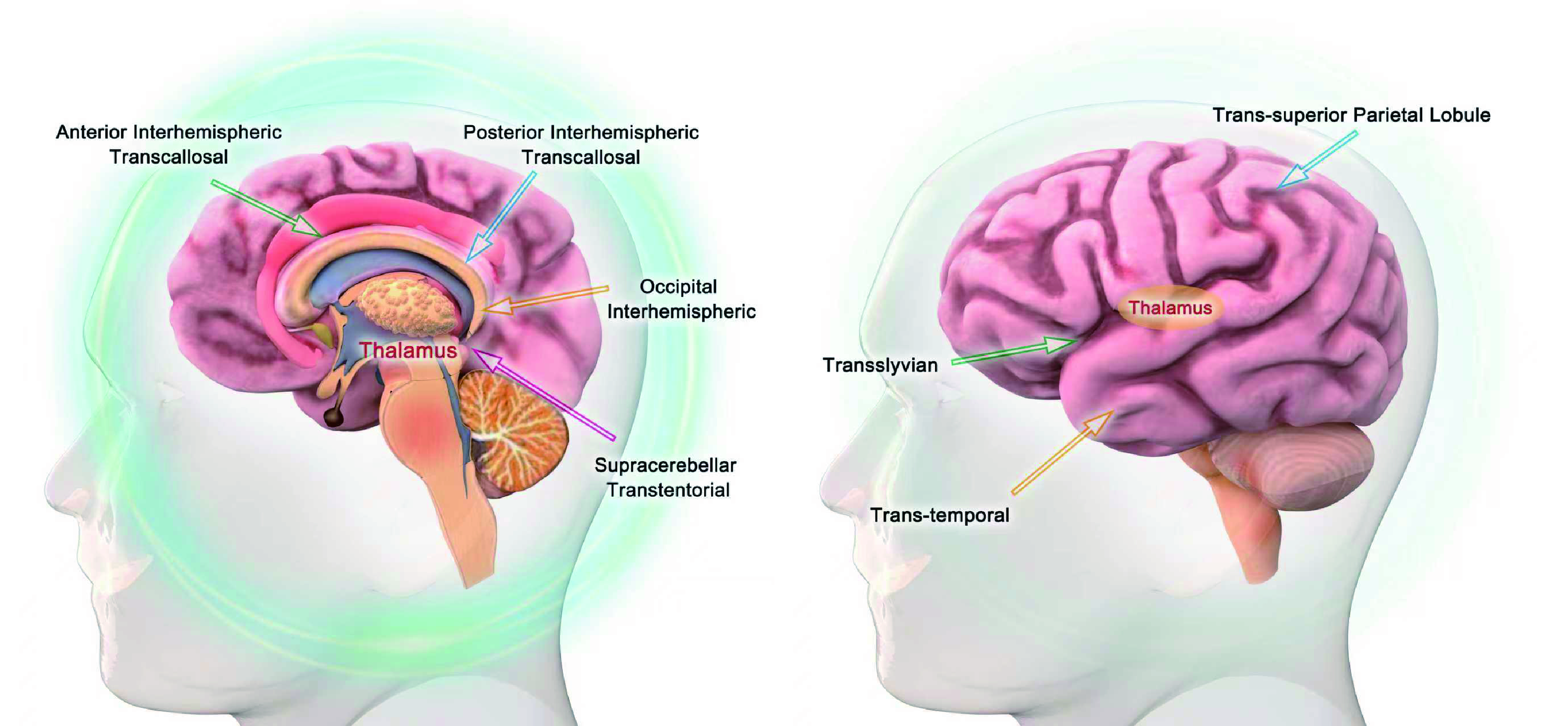
Combining all included studies, the presenting symptoms were diverse, including raised ICP (669 patients), movement disorders (688 patients), sensory disorders (688 patients), visual disorders (566 patients), dysphasia (358 patients), mental changes (337 patients), hydrocephalus (401 patients), seizures (250 patients), and other symptoms (urinary incontinence, memory loss, etc.) (448 patients). The overall incidence of preoperative symptoms was calculated by a random-effects model. The pooled overall incidence rates were 0.52 (95% CI: 0.40-0.95) for raised ICP (Figure 2A), 0.48 (95% CI: 0.39-0.57) for movement disorder (Figure 2B), 0.23 (95% CI: 0.18-0.28) for sensory disorder (Figure 2C), 0.17 (95% CI: 0.10-0.23) for visual disorder (Figure 2D), 0.07 (95% CI: 0.03-0.10) for dysphasia (Figure 2E), 0.27 (95% CI: 0.15-0.39) for mental change (Figure 2F), 0.35 (95% CI: 0.13-0.57) for hydrocephalus (Figure 2G), 0.05 (95% CI: 0.02-0.07) for seizures (Figure 2H), and 0.14 (95% CI: 0.08-0.21) for other symptoms (Figure 2I). A summary of the overall effects of preoperative symptoms was presented in Table 2. A total of 691 patients with thalamic gliomas underwent surgical resection, with detailed surgical approach data available for 620 patients. Of these, 455 patients (73.5%) underwent a lateral approach (transcortical/transventricular, transsylvian, or subtemporal), 139 patients (22.4%) underwent a medial approach (interhemispheric transcallosal), and 26 patients (4.1%) underwent a posterior approach (supracerebellar infratentorial or transoccipital). The commonly used surgical approaches for thalamic tumors are summarized in Figure 3. All 691 patients reported the items about EORs, 585 patients (84.7%) achieved GTR/STR, while 106 patients (15.3%) achieved PR.
Figure 2. Forest plots of the overall incidence of preoperative symptoms. Forest plots of: raised ICP (A), movement disorder (B), sensory disorder (C), visual disorder (D), dysphasia (E), mental change (F), hydrocephalus (G), seizure (H), other symptoms (I).
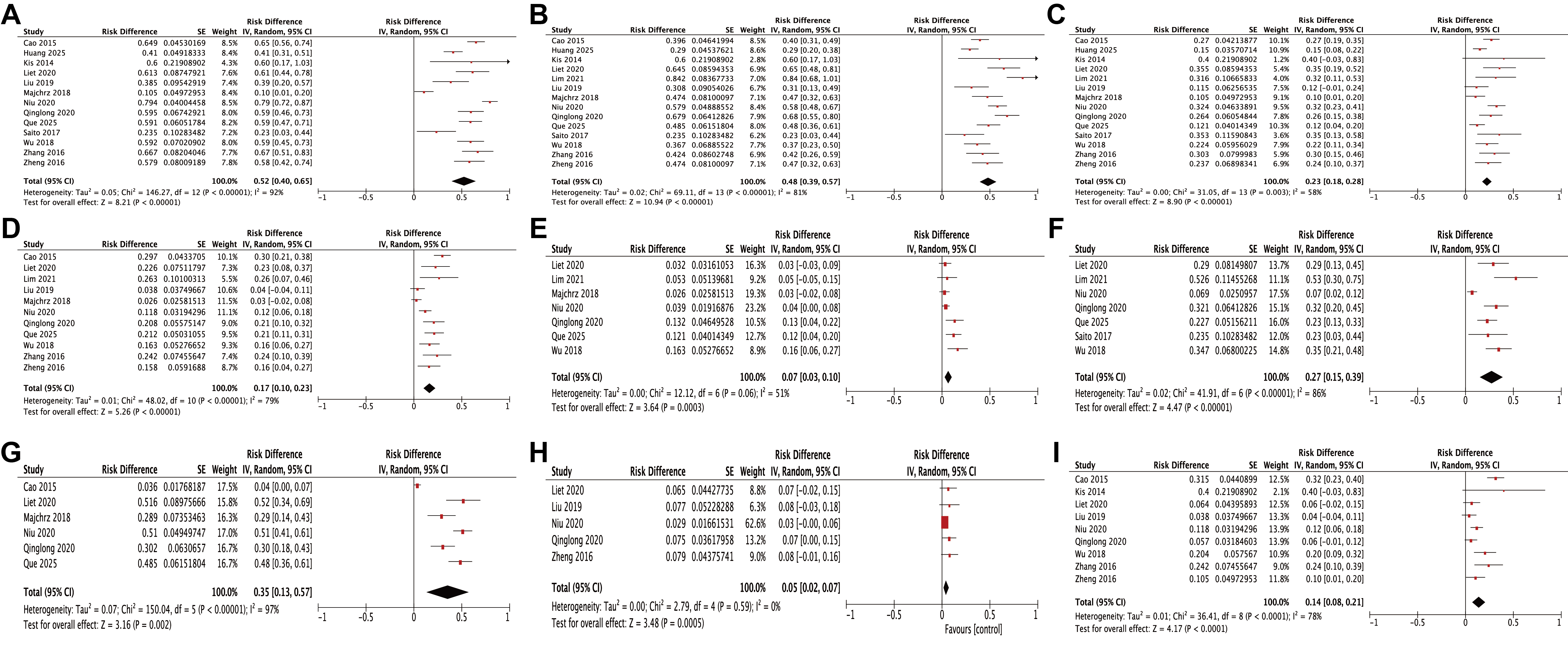
Table 2. The overall effects of preoperative symptoms by Random-Effect Model.
| Preoperative complications | Number of studies included | I² | Overall effect | 95% CI | P value |
|---|---|---|---|---|---|
| Raised ICP | 13 | 92% | 0.52 | 0.40-0.65 | < 0.00001 |
| Movement disorder | 14 | 81% | 0.48 | 0.39-0.57 | < 0.00001 |
| Sensory disorder | 14 | 58% | 0.23 | 0.18-0.28 | < 0.00001 |
| Visual disorder | 11 | 79% | 0.17 | 0.10-0.23 | < 0.00001 |
| Dysphasia | 7 | 51% | 0.07 | 0.03-0.10 | 0.0003 |
| Mental change | 7 | 86% | 0.27 | 0.15-0.39 | < 0.00001 |
| Hydrocephalus | 6 | 97% | 0.35 | 0.13-0.57 | 0.002 |
| Seizure | 5 | 0 | 0.05 | 0.02-0.07 | 0.0005 |
| Other symptoms | 9 | 78% | 0.14 | 0.08-0.21 | < 0.0001 |
Figure 3. Summary of commonly used surgical approaches for thalamic tumors in clinical practice.
Postoperative adjuvant therapy data were available for 323 patients, of whom 72 (22.3%) received radiotherapy alone, 71 (22.0%) received chemotherapy alone, 180 (55.7%) received radiochemotherapy, and 10 (3.1%) did not receive any adjuvant therapy. Li et al.[20], Liu et al.[9], Lim et al.[15], and Saito et al.[18] references or imply Temozolomide-based standard treatment for HGGs/GBM (likely Stupp protocol). Additionally, Saito et al.[18] reported 12 cases of Nimustine use and 1 case of Methotrexate intrathecal administration. Other studies mention radiochemotherapy or chemotherapy without detailing medications, dosages, or schedules. A comprehensive summary of the patients’ treatment modalities is presented in Table 3.
Table 3. The summary of the patients' clinical treatments
| Surgical approach, n=620 | ||
|---|---|---|
| Lateral | Transcortical/Transventricular | 430 (69.4%) |
| Transsylvian | 19 (3.1%) | |
| Subtemporal | 6 (1.0%) | |
| Medial | Interhemispheric transcallosal | 139 (22.4%) |
| Posterior | Supracerebellar-infratentorial | 4 (0.6%) |
| Transoccipital | 22 (3.5%) | |
| EOR, n=691 | ||
| GTR/STR | 585 (86.1%) | |
| PR | 106 (13.9%) | |
| Adjuvant therapy, n=323 | ||
| Radiotherapy | 72 (22.3%) | |
| Chemotherapy | 71 (22.0%) | |
| Radiochemotherapy | 180 (55.7%) | |
| None | 10 (3.1%) | |
Assessment of postoperative complications.
Across all included studies, a spectrum of common postoperative complications was identified, including movement disorder (550 patients), sensory disorder (289 patients), visual disorder (426 patients), language disorder (205 patients), hemorrhage (216 patients), hydrocephalus (504 patients), infection and fever (125 patients), cognitive disorder (315 patients), and other disorders (mutism, electrolyte disturbances, etc.) (233 patients). The overall incidence of complications was calculated using a random-effects model. The pooled overall incidence rates were 0.29 for movement disorders (95% CI: 0.20-0.39) (Figure 4A), 0.13 for sensory disorders (95% CI: 0.09-0.17) (Figure 4B), 0.03 for visual disorders (95% CI: 0.02-0.05) (Figure 4C), 0.09 for language disorders (95% CI: 0.02-0.16) (Figure 4D), 0.09 for hemorrhage (95% CI: 0.05-0.13) (Figure 4E), 0.06 for hydrocephalus (95% CI: 0.03-0.09) (Figure 4F), 0.25 for infection and fever (95% CI: 0.14-0.36) (Figure 4G), 0.06 for cognitive disorder (95% CI: 0.03-0.10) (Figure 4H), and 0.14 for other disorders (95% CI: 0.03-0.25) (Figure 4I). A comprehensive summary of the overall incidence estimates for these complications is presented in Table 4.
Figure 4. Forest plots of the overall incidence of postoperative complications. Forest plots of movement disorder (A), sensory disorder (B), visual disorder (C), language disorder (D), hemorrhage (E), hydrocephalus (F), infection and fever (G), cognitive disorder (H), other disorders (I).
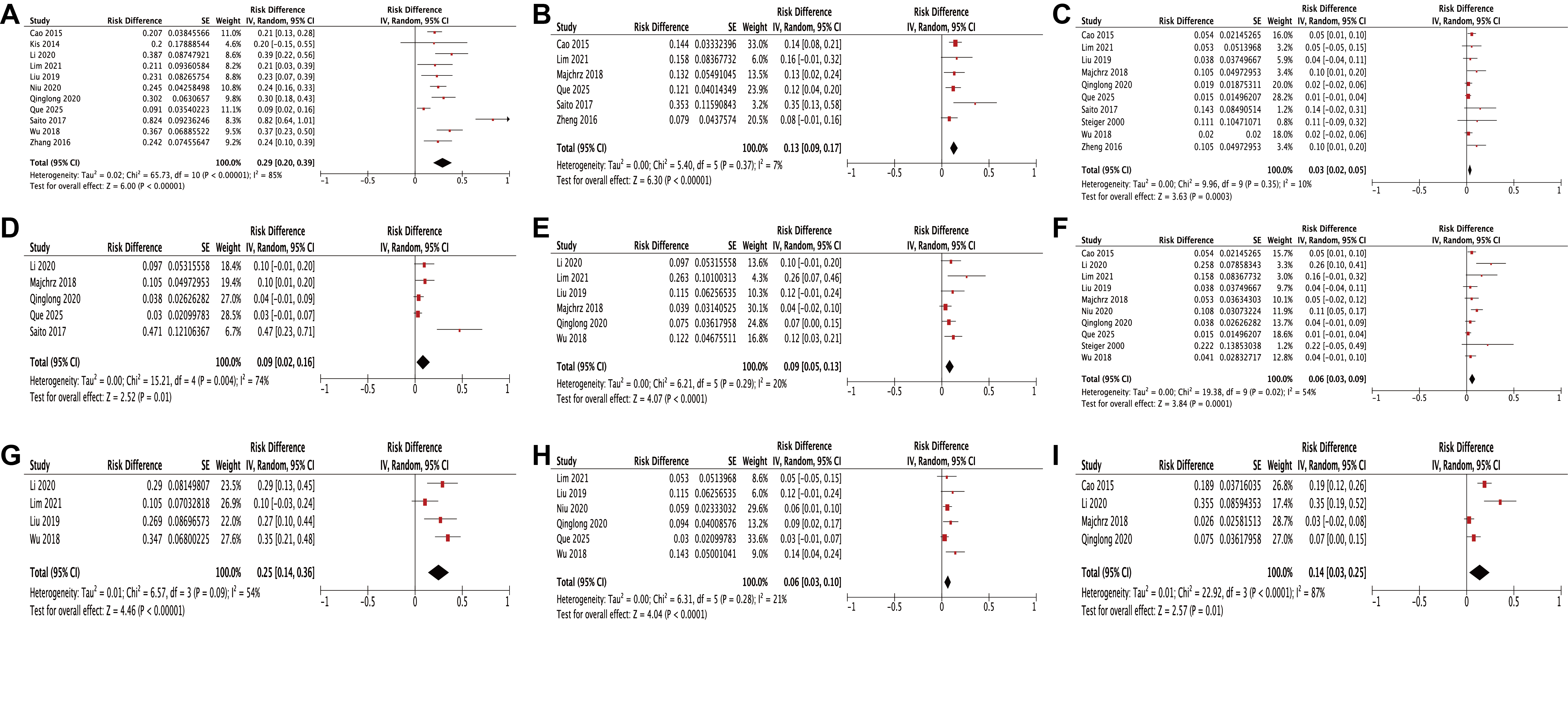
Table 4. The overall effects of postoperative complications by Random-Effect Model.
| Postoperative complications | Number of studies included | I² | Overall effect | 95% CI | P value |
|---|---|---|---|---|---|
| Movement disorder | 11 | 85% | 0.29 | 0.20-0.39 | < 0.00001 |
| Sensory disorder | 6 | 7% | 0.13 | 0.09-0.17 | < 0.00001 |
| Visual disorder | 10 | 10% | 0.03 | 0.02-0.05 | 0.0003 |
| Language disorder | 5 | 74% | 0.09 | 0.02-0.16 | 0.01 |
| Hemorrhage | 6 | 20% | 0.09 | 0.05-0.13 | < 0.00001 |
| Hydrocephalus | 10 | 54% | 0.06 | 0.03-0.09 | 0.0001 |
| Infection and fever | 4 | 54% | 0.25 | 0.14-0.36 | < 0.00001 |
| Cognitive disorder | 6 | 21% | 0.06 | 0.03-0.10 | < 0.0001 |
| Other disorders | 4 | 87% | 0.14 | 0.03-0.25 | 0.01 |
The Cox proportional hazards analysis of the pooled cohort.
We constructed a pooled cohort consisting of 103 patients with a mean age of 41.3 ± 14.5 years and a median OS of 17 months. The median OS for HGGs was 16.1 months, and for LGGs was 23 months. Univariate and multivariate Cox proportional hazards analyses were performed on the pooled cohort to estimate HRs for OS. The variables included in the analyses were age, gender, surgical approach, tumor laterality, EOR, WHO grade, and postoperative adjuvant therapy. In the univariate analysis, postoperative adjuvant therapy was significantly associated with prolonged OS. The HRs were as follows: chemotherapy, HR = 0.11 (95% CI: 0.03–0.37, P < 0.001); radiotherapy, HR = 0.14 (95% CI: 0.04–0.47, P = 0.002); and radiochemotherapy, HR = 0.20 (95% CI: 0.06–0.64, P = 0.007). Kaplan-Meier survival analyses demonstrated that postoperative adjuvant therapy was significantly associated with improved OS (Figure 5G, log-rank test; P = 0.001). No significant associations with OS were observed for the remaining variables (age, gender, surgical approach, tumor laterality, EOR and WHO grade) in the univariate analysis, as illustrated by Kaplan-Meier survival curves (Figures 5A–5F). In the multivariate analysis, postoperative adjuvant therapy continued to demonstrate a significant association with prolonged OS, with HRs of 0.19 for chemotherapy (95% CI: 0.04–0.81, P = 0.025), 0.10 for radiotherapy (95% CI: 0.02–0.42, p = 0.002), and 0.13 for radiochemotherapy (95% CI: 0.03–0.51, p = 0.003). LGGs were significantly associated with prolonged OS, with an HR of 0.34 (95% CI: 0.16–0.73, p = 0.005), a finding not observed in the univariate analysis. The overall Kaplan-Meier survival curve is presented in Figure 5H, and detailed results of the univariate and multivariate Cox analyses are summarized in Table 5.
Figure 5. Kaplan Meier survival curves of each variable in the pooled cohort: age (A), gender (B), surgical approach (C), tumor side (D), EOR (E), WHO grade (F), adjunct therapy (G) and overall OS in pooled cohort (H).
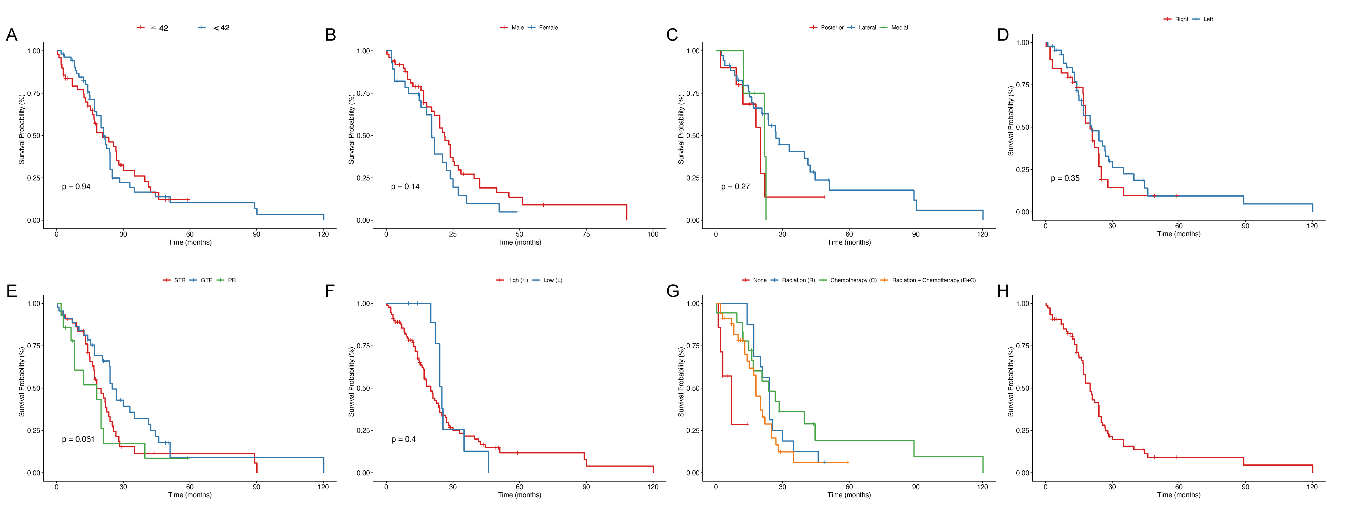
Table 5. Results of univariate and multivariate cox proportional hazard analysis in pooled cohort.
| Cox Univariate | Cox Multivariate | ||||
|---|---|---|---|---|---|
| N | Hazard ratio (95%CI) | P value | Hazard ratio (95%CI) | P value | |
| Age, n=103 | |||||
| ≥42 | 49 | 0.98 (0.97-1) | 0.054 | 0.99 (0.97-1.01) | 0.231 |
| <42 | 54 | - | - | ||
| Gender, n=78 | |||||
| Male | 28 | 1.5 (0.88-2.56) | 0.138 | - | |
| Female | 50 | - | - | ||
| Approach, n=49 | |||||
| Lateral | 35 | 0.53 (0.22-1.26) | 0.15 | - | |
| Medial | 4 | 1 (0.26-3.93) | 0.995 | - | |
| Posterior | 10 | - | - | ||
| Side, n=84 | |||||
| Left | 45 | 0.77 (0.46-1.31) | 0.342 | 1.11 (0.58-2.14) | 0.748 |
| Right | 39 | - | - | ||
| EOR, n=103 | |||||
| GTR | 45 | 0.62 (0.37-1.02) | 0.062 | 0.55 (0.28-1.10) | 0.090 |
| STR | 44 | - | - | ||
| PR | 14 | 1.27 (0.64-2.53) | 0.487 | 1.93 (0.87-4.26) | 0.106 |
| WHO grade, n=103 | |||||
| L | 12 | 0.74 (0.35-1.54) | 0.4 | 0.34 (0.16-0.73) | 0.005 |
| H | 91 | - | - | ||
| Adjunct therapy, n=75 | |||||
| C | 18 | 0.11 (0.03-0.37) | <0.001 | 0.19 (0.04-0.81) | 0.025 |
| R | 16 | 0.14 (0.04-0.47) | 0.002 | 0.10 (0.02-0.42) | 0.002 |
| R+C | 34 | 0.2 (0.06-0.64) | 0.007 | 0.13 (0.03-0.51) | 0.003 |
| None | 7 | - | - | ||
Discussion
This systematic review and meta-analysis provide a comprehensive evaluation of surgical outcomes and complications associated with adult thalamic gliomas, highlighting the complexities of managing these rare and challenging tumors. Across 15 studies (n = 695), GTR or STR achieved in 86.1% of cases , with high-grade gliomas comprising the majority (77.8%)[17, 29]. Although surgical resection remains the cornerstone of management, the considerable incidence of postoperative complications—particularly movement disorders (29%) and infection or fever (25%)—underscores the delicate balance between oncologic control and neurological preservation[13, 15]. Multivariate analysis confirmed that adjuvant therapies (chemotherapy, radiotherapy, or combined radiochemotherapy) significantly prolonged survival, while low-grade histology was associated with better outcomes. These findings highlight the importance of integrating maximal safe resection with multimodal adjuvant therapy to optimize both survival and functional outcomes in this challenging tumor entity.
The predominance of HGGs (77.8%) in our cohort is consistent with the aggressive nature of thalamic gliomas reported in the literature[30]. HGGs are associated with poorer prognosis due to their infiltrative growth and resistance to adjuvant therapies[31]. Our Cox proportional hazards analysis revealed that postoperative adjuvant therapies, including chemotherapy, radiotherapy, and combined radio-chemotherapy, significantly prolonged OS, with HRs of 0.19, 0.10, and 0.13, respectively, in multivariate analysis. These findings corroborate studies demonstrating the synergistic effects of multimodal adjuvant treatments in improving survival outcomes for HGGs[32, 33]. Notably, LGGs were associated with a significantly prolonged OS (HR = 0.34), suggesting that tumor biology plays a pivotal role in determining prognosis[34]. In this study, the detection rate of H3K27M mutation in adult thalamic gliomas was 20.6%, suggesting that this mutation is an important molecular marker for thalamic midline gliomas and is associated with a significantly shortened OS of approximately 9-12 months. According to the 2021 WHO CNS5 classification, H3K27M mutation has been redefined as "H3K27-altered diffuse midline glioma," encompassing alternative events such as EZHIP overexpression or EGFR mutations, further expanding its molecular spectrum[4]. This epigenetic dysregulation promotes treatment resistance, underscoring the need for targeted therapies (e.g., EZH2 inhibitors) and immunotherapies in future trials[35-37]. Multicenter collaboration is needed to establish standardized diagnostic and treatment pathways for this mutation and accelerate the transition of translational research into clinical practice.
The selection of surgical approaches for thalamic lesions should be guided by both tumor-specific anatomical factors and functional considerations. In our cohort, the variability in approaches - lateral (51.8%), medial (25.9%), and posterior (22.3%) - reflects the need to tailor strategies based on lesion location, as demonstrated by Rangel-Castilla and Spetzler's six-region classification system[38]. Lateral approaches, which were most common, may offer better access to tumors extending to the ventricular or cortical surfaces, potentially reducing morbidity compared to medial or posterior approaches that traverse deeper structures[39]. While lateral approaches were most common, particularly for tumors extending to ventricular or cortical surfaces, our analysis found no significant association between surgical approach and overall survival. This supports the prevailing view that EOR is a more critical prognostic factor than the specific surgical corridor employed[40, 41]. The high rate of GTR/STR in our cohort suggests that advancements in intraoperative technologies, such as neuronavigation and tractography, have enhanced surgical precision, minimizing damage to eloquent areas[42, 43]. For anteroinferior lesions, the orbitozygomatic approach minimizes internal capsule risk, while the anterior contralateral transcallosal route optimizes exposure for lateral lesions through gravity-assisted retraction. Posterior lesions may benefit from supracerebellar-infratentorial approaches that leverage natural anatomical corridors. These findings underscore that while anatomical considerations should drive approach selection, the primary surgical goal remains maximal safe resection, facilitated by advanced intraoperative guidance systems. Future refinements may incorporate emerging techniques like laser interstitial thermal therapy for select deep-seated lesions, though open microsurgical approaches currently remain the gold standard for thalamic tumor resection.
Postoperative complications remain a significant concern, with movement disorders being the most frequent (29%), likely due to the thalamus’s proximity to the corticospinal tracts[44, 45]. The relatively high incidence of infection and fever (25%) may be attributed to prolonged surgical times and the use of intraoperative monitoring devices, which increase the risk of nosocomial infections[46]. Hydrocephalus (6%) and hemorrhage (9%) were less frequent but still notable, highlighting the need for meticulous surgical planning and postoperative care[47]. These complications align with reports on deep-seated brain tumor surgeries, where the risk of neurological deficits is amplified by the thalamus’s complex neuroanatomy[48]. It is also influenced by patient factors (e.g., frailty, H3K27M status), and emerging technologies such as augmented reality navigation and machine learning risk models are expected to improve the prediction and prevention of complications.
The heterogeneity in tumor histology and patient demographics across studies complicates direct comparisons, a limitation acknowledged in prior meta-analyses[49]. The lack of standardized definitions for EOR and surgical approaches further challenges the generalizability of our findings[50]. Future studies should aim to standardize these parameters and incorporate molecular profiling, such as IDH, H3K27M mutation status, which has been shown to influence prognosis in gliomas[35, 51]. Additionally, the integration of advanced imaging modalities, like functional MRI and diffusion tensor imaging, could further refine surgical strategies by mapping critical neural pathways[52, 53].
Our findings highlight the critical importance of a multidisciplinary team (MDT) approach in managing thalamic tumors, where the integration of maximal safe resection with tailored adjuvant therapies (chemotherapy and radiotherapy) has demonstrated significant survival benefits[54]. The MDT discussion should specifically address: (1) molecular profiling (e.g., H3K27M status) to guide targeted therapy selection, (2) radiation field optimization to minimize cognitive sequelae while ensuring adequate tumor coverage, and (3) sequential treatment planning to balance efficacy and toxicity, particularly for patients with pre-existing neurological deficits. Recent advances in immunotherapy and molecular targeted agents (e.g., EZH2 inhibitors for H3K27M-mutant tumors) should be incorporated into MDT deliberations, with consideration for clinical trial enrollment when appropriate. This collaborative decision-making process is essential to optimize the risk-benefit ratio of combined modality therapy while addressing the unique anatomical and biological challenges posed by thalamic tumors.
This meta-analysis has several limitations. First, heterogeneity in study designs, patient demographics, and surgical techniques may affect result generalizability. Second, inconsistent definitions of outcomes and reporting biases in retrospective studies could influence pooled analyses. Third, limited molecular profiling data (e.g., IDH, H3K27M status) restricts prognostic assessments. Fourth, the exclusion of non-English studies and potential publication bias may skew findings. Finally, variability in follow-up durations and adjuvant therapy details complicates long-term outcome evaluations. These limitations highlight the need for standardized reporting and prospective studies to validate conclusions.
Conclusion
This meta-analysis demonstrates that maximal safe resection combined with adjuvant therapy significantly improves survival in adult thalamic gliomas, despite high complication rates. The findings support tailored surgical strategies guided by tumor location and advanced intraoperative technologies to minimize morbidity. While adjuvant therapies—particularly chemotherapy and radiotherapy—enhance outcomes, the high prevalence of H3K27M mutations underscores the need for targeted treatments. Standardized reporting of surgical approaches, molecular profiling, and long-term follow-up are essential to refine management. Future research should focus on integrating precision medicine and novel therapies to further optimize survival and functional preservation in these challenging tumors.
Abbreviations
AA - Anaplastic Astrocytoma; AOA - Anaplastic Oligoastrocy-toma; CI - Confidence Interval; CNS - Central Nervous System; DA - Diffuse Astrocytoma; DMG - Diffuse Midline Glioma; EOR - Extent of Resection; FA - Fibrillary Astrocytoma; GA - Gemisto-cytic Astrocytoma; GBM - Glioblastoma Multiforme; GG - Gan-glioglioma; GSM - Gliosarcoma; GTR - Gross Total Resection; HGG - High-Grade Glioma; HR - Hazard Ratio; ICP - Intracranial Pressure; IDH - Isocitrate Dehydrogenase; LGG - Low-Grade Glioma; MDT - Multidisciplinary Team; MeSH - Medical Subject Headings; MGMT - O⁶-Methylguanine-DNA Methyltransferase; NR - Not Reported; OA - Oligoastrocytoma; OS - Overall Surviv-al; PCA - Pilocytic Astrocytoma; PR - Partial Resection; PRIS-MA - Preferred Reporting Items for Systematic Reviews and Meta-Analyses; PXA - Pleomorphic Xanthoastrocytoma; SD - Standard Deviation; STR - Subtotal Resection; WHO - World Health Organization.
Declarations
Author Contributions
Conceptualization: Hongliang Mao, Fengchun Mu, Xinyu Wang, Jinghai Wan, Ming Yang. Data curation: Hongliang Mao, Fengc-hun Mu. Formal analysis: Hongliang Mao, Fengchun Mu, Xinyu Wang, Jinghai Wan, Ming Yang. Funding acquisition: Jinghai Wan, Ming Yang. Investigation: Hongliang Mao, Fengchun Mu, Xinyu Wang. Methodology: Hongliang Mao, Fengchun Mu, Xinyu Wang, Jinghai Wan, Ming Yang. Project administration:Ming Yang. Resources: Hongliang Mao, Fengchun Mu, Ming Yang Supervision: Jinghai Wan, Ming Yang. Validation: Feng-chun Mu, Jinghai Wan, Ming Yang. Writing—original draft: Hongliang Mao, Fengchun Mu. Writing—review & editing: Jing-hai Wan, Ming Yang.
Acknowledgements
We appreciate the efforts of our colleagues to cooperate and complete this work, and we also thank the support by the Pro-fessor Ming Yang in this work.
Funding information
This work was funded by CAMS Innovation Fund for Medical Sciences (2024-I2M-3-014, 2022-I2M-C&T-B-063), the Na-tional Natural Science Foundation of China (No. 82103231, No.82072803 and No.82472722) and Beijing Hope Run Special Fund of Cancer Foundation of China (No. LC2022B18).
Ethics Approval and Consent to Participate
This systematic review and meta-analysis did not require eth-ical approval as it synthesized data from previously published studies. All included studies were conducted in accordance with their respective institutional ethical standards.
Competing Interests
The authors declare no conflicts of interest.
Data availability
The data that support the findings of this study are available from the corresponding author upon reasonable request.
References
 Figures
 References
 Peer
Peer Information
Figure 1. Flowchart of studies that could be quantitatively synthesized according to the inclusion and exclusion criteria.
Figure 2. Forest plots of the overall incidence of preoperative symptoms. Forest plots of: raised ICP (A), movement disorder (B), sensory disorder (C), visual disorder (D), dysphasia (E), mental change (F), hydrocephalus (G), seizure (H), other symptoms (I).
Figure 3. Summary of commonly used surgical approaches for thalamic tumors in clinical practice.
Figure 4. Forest plots of the overall incidence of postoperative complications. Forest plots of movement disorder (A), sensory disorder (B), visual disorder (C), language disorder (D), hemorrhage (E), hydrocephalus (F), infection and fever (G), cognitive disorder (H), other disorders (I).
Figure 5. Kaplan Meier survival curves of each variable in the pooled cohort: age (A), gender (B), surgical approach (C), tumor side (D), EOR (E), WHO grade (F), adjunct therapy (G) and overall OS in pooled cohort (H).
Peer-review Terminology
Identity transparency: Single anonymized
Reviewer interacts with: Editor
Details
This is an open access article under the terms of the Creative Commons Attribution License(http://creativecommons.org/licenses/by/4.0/), which permits use, distribution and reproduction in any medium, provided the original work is properly cited.
Publication History
Received 2025-10-13
Accepted 2025-11-05
Published 2025-11-22