




Abstract
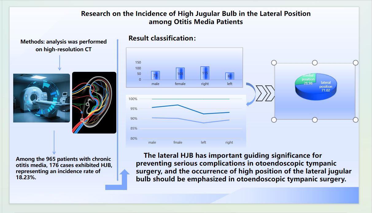
Objective:This study aims to analyze the incidence of high jugular bulb (HJB), an anatomical variation, in patients with otitis media using high-resolution CT imaging in the lateral position.
Methods:A retrospective analysis was performed on high-resolution CT scans of the middle ear from 965 patients clinically diagnosed with otitis media between October 2018 and October 2023. The age range was 6 to 81 years, with a mean age of 47.11 years. The cohort included 387 male and 578 female patients. HJB was defined as the superior border of the jugular bulb extending beyond the inferior border of the basal turn of the cochlea. Using the lateral wall of the cochlear basal turn as the reference line, HJB was classified into medial and lateral types. In the lateral type, the jugular bulb was in proximity to the tympanic membrane, ossicular chain, or round window; in the medial type, it was adjacent to the cochlear aqueduct, vestibular aqueduct, or internal auditory canal. The presence of HJB was recorded, and gender and side distribution were analyzed.
Results:Among the 965 patients with chronic otitis media, 176 cases exhibited HJB, representing an incidence rate of 18.23%. Of these, 103 were female (58.52%) and 73 were male (41.48%), indicating a higher prevalence in females. Right-sided HJB occurred in 115 cases (65.34%), compared to 61 cases on the left (34.66%), suggesting a predominance on the right side. Lateral HJB was observed in 125 cases (71.02%), including five cases with direct exposure, whereas medial HJB was found in 51 cases (28.98%), demonstrating a significantly higher frequency of the lateral variant.
Conclusions:The findings indicate that HJB is more prevalent in females than males and more common on the right side than the left. Additionally, the lateral type occurs more frequently than the medial type. Given its anatomical significance, lateral HJB must be carefully evaluated during otoscopic tympanic surgery to prevent potentially serious complications. Special attention should be paid to identifying this variation preoperatively.
Keywords:Otitis media; High jugular bulb; Otoscope; Tympanic surgery
Introduction
Chronic otitis media is a prevalent condition within the field of otorhinolaryngology. In recent years, as scientific knowledge has become more widespread, public awareness regarding the prevention and treatment of otitis media has improved significantly. Consequently, there has been a notable decrease in the number of patients suffering from severe otitis media or extensive lesions, with the majority now presenting with simple otitis media characterized by tympanic membrane perforation. As a result, the number of patients requiring traditional mastoidectomy has diminished, leading to a rapid advancement in otoscopic technology. Currently, minimally invasive otoscopic surgery has emerged as the primary surgical approach for chronic otitis media. The high jugular bulb (HJB) is a common anatomical variation of the temporal bone encountered in clinical practice, which holds considerable importance for otological surgery[1]. Notably, the lateral HJB variation can significantly impact the execution of otoscopic procedures. In particular, when there is a defect or thinning of the bone wall at the top or lateral aspect of the jugular bulb, the risk of complications during surgery increases. Inadequate management during the procedure can result in severe bleeding and may even pose a threat to the patient's life, often necessitating the termination of the surgery[2]. Therefore, accurately diagnosing HJB anatomical variations prior to otoscopic surgery is crucial for anticipating potential risks and challenges associated with the operation.
This report presents a retrospective analysis of HJB variations in patients with chronic suppurative otitis media, as identified through high-resolution CT scans of the middle ear, along with an assessment of the likelihood of high-position variations in the lateral position.
Materials and Methods
General InformationA retrospective study was carried out involving 965 patients diagnosed with chronic suppurative otitis media who underwent high-resolution CT scans of the middle ear at the otorhinolaryngology-head and neck surgery outpatient department of Bozhou People's Hospital between October 2018 and October 2023. This study received approval from the Ethics Committee of Bozhou City People's Hospital and adheres to the principles outlined in the Helsinki Declaration. The ages of the patients ranged from 6 to 81 years, with an average age of 47.11 years.
Scanning MethodFor the middle ear high-resolution thin-layer scanning, all patients received standard axial and coronal plain scans of the temporal bone. The scanning baseline was set at the orbitomeatal line, with a slice thickness of 0.5 to 1.0 mm, a matrix size of 512×512, and a range of /L: 4000/400 Hu to 800 Hu. Scanning was conducted continuously from 5 mm below the baseline to the upper edge of the petrous pyramid, followed by MPR coronal and sagittal three-dimensional reconstructions for assessment.
Diagnostic CriteriaThe diagnosis of a high jugular bulb (HJB) was determined by the criterion that the upper edge of the jugular bulb was positioned above the lower edge of the cochlea's basal turn. The HJB was classified into medial high-position and lateral high-position based on the lateral wall of the cochlea's basal turn as a boundary. In the lateral high-position, the bulb was in contact with the tympanic membrane, ossicular chain, or round window, while in the medial high-position, it was in contact with the cochlear aqueduct, vestibular aqueduct, and internal auditory canal[15].
Statistical MethodData analysis was performed using SPSS Statistics 20.0 software. For categorical data, the chi-square test was utilized for inter-group comparisons, with a significance level set at P < 0.05 to indicate statistically significant differences.
Results
The basic characteristics of patients with chronic otitis mediaAmong the 965 patients with chronic otitis media included in the investigation and analysis, 176 patients had HJB, accounting for 18.23% of the total number of otitis media patients. Among the patients with HJB, there were 103 female patients, accounting for 58.52% of the total positive cases, and 73 male patients, accounting for 41.48%, with a higher proportion in females than in males. There were 115 cases of high - position on the right side, accounting for 65.34% of the total positive cases, and 61 cases of high - position on the left side, accounting for 34.66%, with a higher proportion on the right side than on the left side. There were 125 cases of lateral high - position, accounting for 71.02% of the total positive cases, among which 5 cases were exposed, and 51 cases were of medial high - position, accounting for 28.98%, with a higher incidence of lateral high - position than medial high - position.
The comparison of gender and left - right sides is shown in Table 1 below, and the difference was statistically significant.
Table 1 The comparison of gender and left - right sides
| right high position | left high position | |
|---|---|---|
| male | 54 | 19 |
| female | 61 | 42 |
| χ2 | 4.104 | |
| p | 0.043 |
The comparison of genders in lateral high - position and medial high - position is shown in Table 2 below, and the difference was statistically significant.
Table 2 The comparison of genders in lateral high-position and medial high-position
| male | female | |
|---|---|---|
| Lateral position HJB | 45 | 80 |
| Medialis HJB | 28 | 23 |
| p | 5.331 | |
| χ2 | 0.021 |
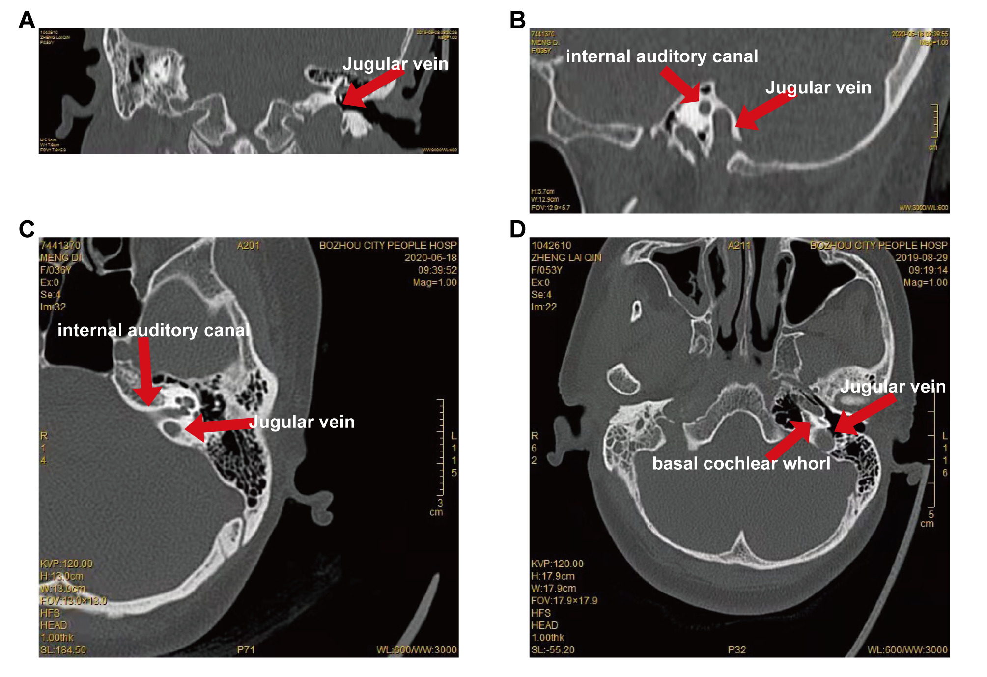
Imaging Pictures of Typical Cases We listed some typical pictures to deepen the understanding of this disease. Such as thinning change of the lateral high - position of the jugular bulb (Fig. 1A), medial high - position change of the jugular bulb (Fig. 1B and 1C) and exposure change of the lateral high - position of the jugular bulb (Fig. 1D).
Figure 1. Imaging Pictures of Typical Cases.
A Thinning Change of the Lateral High - position of the Jugular Bulb.
The bone is thin on the lateral side of the jugular vein bulb.
Discussion
The jugular bulb is situated in the jugular fossa beneath the petrous bone. A high jugular bulb (HJB) is a frequent vascular variation, with most instances being asymptomatic[3]. The reported prevalence of HJB, defined as a high-position jugular bulb that reaches or surpasses the level of the basal turn of the cochlea, ranges from 6% to 24%. In patients with chronic otitis media, the incidence is 20.3%. Ears affected by chronic otitis media are often more likely to have HJB, which is relatively common[4]. This study found an incidence of 18.23%, supporting previous findings. Additionally, the right internal jugular bulb was found to be higher than the left, possibly due to a right-sided dominance in venous vessels, as the right jugular bulb is typically larger. The occurrence of HJB was also higher in women than in men, potentially linked to estrogen levels, aligning with earlier research[5-7]. Furthermore, the analysis revealed that lateral HJB was more prevalent among otitis media patients compared to medial HJB, with some patients exhibiting lateral high-position alongside exposure.
HJB can exhibit three morphological variations: (1) A defect in the upper and lateral walls of the high-position jugular fossa, allowing the jugular bulb to protrude into the middle ear; (2) An incomplete bony covering of the high-position jugular bulb and its fossa; (3) The high-position jugular bulb and its fossa extending into the tympanic cavity from below, covered by a thin, complete bony plate[8]. Different locations and orientations of the high-position jugular bulb can lead to varying symptoms[9 - 14]. The jugular bulb can be categorized into medial high-position and lateral high-position based on its relation to the lateral wall of the basal turn of the cochlea. In the lateral high-position, the bulb contacts the tympanic membrane, ossicular chain, or round window, while in the medial high-position, it contacts the cochlear aqueduct, vestibular aqueduct, and internal auditory canal[15]. The position of HJB in the hypotympanum can be classified into two types: (1) The anterior-inferior quadrant of the tympanic isthmus; (2) The posterior-inferior quadrant of the tympanic isthmus. Both scenarios pose a risk of damaging the jugular bulb during middle-ear surgery, with the former often being injured during the dissection of inflammatory granulation tissue and the latter during the elevation of the tympanomeatal flap, which can tear the jugular bulb[16].When the jugular bulb is positioned laterally and high, with thin surrounding bone, there may be just a soft tissue membrane between it and the surrounding area, and it could even extend into the posterior-inferior tympanum without any protection. In such situations, there is a high risk of injuring the jugular bulb and causing bleeding during ear surgery. Careless handling in this scenario can pose a serious risk to the patient's life[17].
For patients with otitis media who have a high jugular bulb (HJB), it is advisable to refrain from performing blind tympanic exploration, tympanic membrane incision, or blind tympanic membrane puncture[18]. Conducting a pre-operative high-resolution CT scan of the middle ear can help identify the lateral high position of the HJB, allowing otolaryngologists to better understand the patient's unique anatomical features and the specific characteristics of the lesion prior to tympanic surgery. This knowledge aids in selecting the most appropriate surgical approach and being aware of potential intraoperative risks, which can help prevent complications such as excessive bleeding during and after the procedure, as well as serious issues like intracranial hypertension, intracranial hemorrhage, sigmoid sinus thrombosis, and hemiplegia[19 - 20].
Conclusion
Consequently, it is crucial to focus on the lateral high-position HJB variation prior to surgery, as it holds significant importance for otoscopic tympanic procedures.
Declarations
Author contributions
Qingjun Ji(First Author): writing original draft, prepare, create, or express the content for publication, especially in writing the initial draft, including substantive translation. Chunbo Feng: writing review and editing, prepare, create, or express the content for publication, especially in writing the initial draft, including substantive translation. Li Wang:writing review and editing, prepare, create, or express the content for publication, especially in writing the initial draft, including substantive translation. Fuyou Xie:writing review and editing, prepare, create, or express the content for publication, especially in writing the initial draft, including substantive translation. Dapeng Li:supervision, supervise and lead the planning and execution of research activities. Wei Chai (Corresponding Author): supervision, supervise and lead the planning and execution of research activities. All authors read and approved the final manuscript.
Acknowledgements
We would like to express our heartfelt appreciation to the Science and Technology Bureau of Bozhou City and the Research Fund of Bozhou People's Hospital for their invaluable assistance. We also extend our thanks to the Imaging Center of Bozhou People's Hospital for supplying the original image data, Dr. Li Dapeng from the Science and Education Department of Bozhou People's Hospital for his valuable advice on the thesis, and Director Chai Weike from the Department of Otorhinolaryngology Head and Neck Surgery of Bozhou People's Hospital for his steadfast support. Furthermore, we are thankful to Dr. Wang Li and Dr. Feng Chunbo from the same department for their helpful guidance on the thesis.
Funding information
Funding: Key Research and Development Program Project of Bozhou Science and Technology Bureau, Anhui Province in 2023 (Project No.: bzzc2023053).
Ethics approval and consent to participate
Funding: Key Research and Development Program Project of Bozhou Science and Technology Bureau, Anhui Province in 2023 (Project No.: bzzc2023053).
Competing Interests
The authors declare that they have no existing or potential commercial or financial relationships that could create a conflict of interest at the time of conducting this study.
Data availability
The data are available from the corresponding author on reasonable request.
References
 Figures
 References
 Peer
Peer Information
Figure 1. Imaging Pictures of Typical Cases. A Thinning Change of the Lateral High - position of the Jugular Bulb. The bone is thin on the lateral side of the jugular vein bulb. B and C Medial High - position Change of the Jugular Bulb. D Exposure Change of the Lateral High - position of the Jugular Bulb.
Peer-review Terminology
Identity transparency: Single anonymized
Reviewer interacts with: Editor
Details
This is an open access article under the terms of the Creative Commons Attribution License(http://creativecommons.org/licenses/by/4.0/), which permits use, distribution and reproduction in any medium, provided the original work is properly cited.
Publication History
Received 2025-06-13
Accepted 2025-07-16
Published 2025-08-10