




Abstract
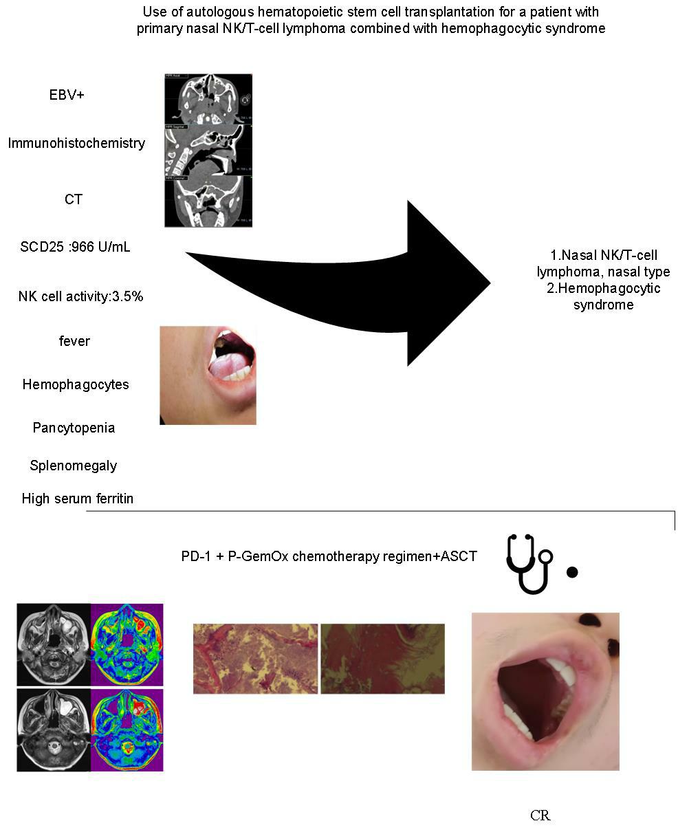
Extranodal NK/T-cell lymphoma, nasal type (ENKTCL-NT) is a type of primary nasal peripheral T-cell lymphoma, which is a rare type of non-Hodgkin's lymphoma, especially with hemophagocytic syndrome (HPS), and can easily lead to clinical misdiagnosis. The disease is dangerous, it progresses rapidly, and the treatment is ineffective. This article reports a case of ENKTCL-NT with HPS, which involves the primary nasal cavity invading the oral cavity. Patients attain improved remission outcomes with autologous hematopoietic stem cell transplantation in combination with chemotherapy. This paper reviews current domestic and international research and summarizes diagnostic and therapeutic approaches to enhance clinicians' comprehension of the disease.
Keywords:Extranodal NK/T-cell lymphoma, hemophagocytic syndrome, autologous hematopoietic stem cell transplantation
Case report
We present a rare case of Extranodal NK/T-cell lymphoma, nasal type (ENKTCL-NT) involving the oral and maxillofacial region with HPS in a 14-year-old male. The patient initially presented with sinusitis-like symptoms but exhibited poor responsiveness to conventional treatment. He was subsequently diagnosed with Epstein-Barr virus (EBV)-positive T-cell proliferative disease at a renowned hospital in Shanghai. Positron emission tomography-computed tomography (PET-CT) revealed increased fluorodeoxyglucose (FDG) uptake in the cervical lymph nodes, and lymph node biopsy suggested lymphoma. ENKTCL-NT was confirmed via immunohistochemical analysis. Within months, the tumor rapidly progresses to involve the oral and maxillofacial region, leading to severe complications, including nasal and oral bleeding, hemorrhagic shock, and HPS. Given the aggressive nature of the disease, a multidisciplinary team consultation was conducted to confirm the diagnosis. The patient achieved complete remission (CR) following standard chemotherapy in combination with autologous hematopoietic stem cell transplantation (ASCT). Free skin grafting, performed by the Department of Stomatology, effectively restored the patient's facial appearance. ENKTCL-NT is a rare malignancy, particularly when accompanied by HPS. Its early clinical manifestations are often nonspecific, frequently leading patients to initially present in otolaryngology departments due to nasal symptoms. This case report details the successful application of chemotherapy combined with ASCT in the treatment of ENKTCL-NT with hemophagocytic syndrome (HPS), offering valuable insights for clinical management.
A patient (hospital ID: 1665984) who was a 15-year-old male was admitted to the Department of Pediatrics at Bozhou People's Hospital on June 4, 2025. Medical history: A patient with a four-day history of fever and cough was admitted to the Department of Pediatrics at Bozhou People's Hospital on November 3, 2023. A coronal CT scan of the paranasal sinuses revealed inflammation in the left frontal sinus, ethmoid sinus, sphenoid sinus, and maxillary sinus. Additionally, hypertrophy of the bilateral inferior turbinates and slight deviation of the nasal septum were noted. Sinusitis was suspected; however, no improvement was observed following anti-infective treatment. The patient was subsequently transferred to a pediatric medical center in Shanghai. There were some atypical lymphocytes in the patient's peripheral blood, and the patient was EBV positive; therefore, he was considered to have EBV-positive T-cell proliferative disease in the pediatric medical center in Shanghai. Further examination and treatment are recommended. However, the family declined this option because it was far from home and not convenient to take care of.
The patient was admitted to the PICU+NICU of our hospital due to oral bleeding on January 8, 2024. The diagnoses included (1) hemorrhagic shock and (2) EBV-positive T-cell proliferative disease in children. Following consultation with the otolaryngology department, tamponade compression was attempted to control the bleeding; however, the patient did not cooperate. Tracheal intubation and ventilator-assisted breathing were then initiated. Physical examination revealed swelling of the nasal mucosa, absence of most of the left soft palate with communication with the nasal cavity, visible blood clots, and significant oozing of blood. After sedation during intubation, further exploration of the oral and nasal cavities revealed no obvious signs of active bleeding. He was considered to have EBV-positive T-cell proliferative disease at the pediatric Medical Center in Shanghai. Therefore, PET-CT imaging is necessary, and PET-CT imaging demonstrated increased FDG metabolism in cervical lymph nodes in January 2024.
On January 22, 2024, nasopharyngeal CT revealed thickening of the soft tissue on the right posterior wall of the nasopharynx, slight narrowing of the right parapharyngeal space, and a soft tissue thickening shadow in the left palate with uneven density. No markedly enlarged lymph nodes were observed adjacent to the carotid sheath bilaterally (Figure 1) . PET-CT combined with basic patient information, lymph node biopsy, and nasal mass biopsy revealed EBV-positive T-cell proliferative disease. Following consultation with the hematology department of our hospital, lymphoma was highly suspected, and immunohistochemical analysis was recommended for further evaluation. During the disease course, the patient presented with recurrent fever, pancytopenia, and splenomegaly. If necessary, serum ferritin, blood lipids, NK cell activity, and soluble CD25 (SCD25) should be measured to determine the presence of HPS. Pathological examination of the nasal mass suggested NK/T-cell lymphoma. On January 17, 2024, immunohistochemistry results of the nasal mass revealed an unclear lymphoid follicular structure, with the following markers: ALK (-), BCL2 (-), BCL6 (-), CD2 (-), CD4 (-), CD8 (-), CD15 (-), CD20 (-), CD38 (-), CD56 (+), epithelial marker (EAM) positive, Ki-67 (85%), LCA (+), MPO (-), PAX5 (-), TDT (-), TIA-1 (+), and in situ EBER (70%+). Based on these findings, the patient was diagnosed with nasal-type NK/T-cell lymphoma (nasopharyngeal lesion). The laboratory results revealed that the SCD25 level was 966 U/mL, the NK cell activity was decreased (3.5%), and the ferritin level was >1675.56 ng/mL. Hemophagocytes were observed in peripheral blood smears. This study was performed in accordance with the latest diagnostic guidelines and standards. The final diagnosis included the following: 1. Nasal NK/T-cell lymphoma, nasal type (Lugano stage IV, NKTCL prognostic score 3, high-risk group)[1]; 2. Oral bleeding; 3. Hemorrhagic shock; 4. Hypoproteinaemia; 5. Hemophagocytic syndrome [6]. Following initial symptomatic management, including anti-infection therapy and volume expansion, patients with poor general conditions and those with HPS cannot tolerate the toxicity of high-intensity regimens. The use of a reduced-dose P-GemOx regimen can reduce the risk of bone marrow suppression and organ damage.
Figure 1. CT of the patient showing thickening of the soft tissue in the right posterior wall of the nasopharynx, mild narrowing of the right parapharyngeal space, and thickening of the soft tissue in the left palate with heterogeneous density. No significant enlargement of the lymph nodes adjacent to the bilateral carotid sheaths was observed. The paranasal sinus mucosa is thickened, and flaky soft tissue density shadows are evident.

The patient underwent a reduced-dose P-GemOx chemotherapy regimen (gemcitabine 1 g on days 1 and 8; oxaliplatin 150 mg on day 1; pegaspargase 3500 IU on day 2), accompanied by active management of hemophagocytic syndrome. On February 16, 2024, the patient received a reduced-dose PD-1 + P-GemOx regimen, consisting of gemcitabine (1 g on days 1 and 8), oxaliplatin (150 mg on day 1), pegaspargase (3500 IU on day 2), and camrelizumab (200 mg on day 4). Hematopoietic stem cell mobilization was initiated on April 22, 2024, using the VP16 regimen (1.6 g/m2, total dose 2.2 g). Peripheral blood stem cells were successfully harvested on May 5, 2024, yielding 200 mL of enriched product with a CD34+ cell count of 2105.28 cells/μl. The reduced-dose PD-1 + P-GemOx chemotherapy regimen was continued on June 3 and July 1, 2024.
Beginning on August 10, 2024, the patient received BeEAM conditioning therapy (IBM: 1.5 m2), including bendamustine (120 mg/m2, total dose 175 mg, administered on days –8 to –7), cytarabine (200 mg/m2, total dose 250 mg, on days –6 to –3), etoposide (200 mg/m2, total dose 250 mg, on days –6 to –3), and melphalan (140 mg/m2, total dose 200 mg, on day –2). Symptomatic and supportive care measures—hydration, alkalinization, and antiemetic therapy—were provided throughout the course. On August 18, 2024, 400 mL of stem cells were reinfused without complications, resulting in successful engraftment. The patient was subsequently discharged. Maintenance therapy with camrelizumab (200 mg every two months) was initiated on October 13, 2024.
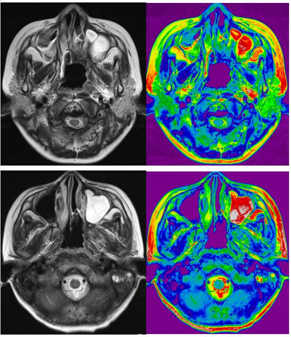
The patient underwent extensive resection of the palatal lesion, free composite tissue transplantation, free skin grafting, full-thickness skin grafting, and tooth extraction at the Department of Stomatology at our hospital on January 8, 2025. A pathological examination of the palate conducted on January 9, 2025, revealed degenerative osteoid tissue and extensive inflammatory necrotic tissue, with no evidence of malignancy (Figure 2). The surgical flap healed successfully without signs of infection. The patient has since shown favorable recovery, with satisfactory reconstruction of the nasal cavity and maxillofacial regions(Figure 3), and the treatment outcome was classified as CR. On March 11, 2025, due to localized swelling, the patient underwent a second resection of the left jaw under local anesthesia. Histopathological analysis of the left maxilla showed extensive infiltration of acute and chronic inflammatory cells within fibrous connective tissue covered by squamous epithelium, accompanied by granulation tissue hyperplasia. The focal areas exhibited mild lymphocytic enrichment, and occasional inflammatory exudates and necrotic-like tissues were observed. No neoplastic cells were identified in the pathological tissue (Figure 4), suggesting stable disease.
Figure 2.Pathological examination of the patient's left mandible revealed the absence of tumor cells.
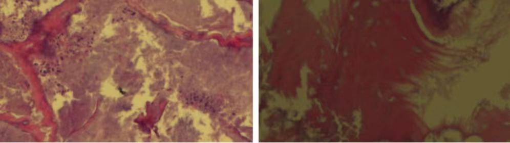
Figure 3. Prior to treatment, the patient's ulcerated maxilla exhibited communication with the nasal cavity, accompanied by a significant amount of yellow secretions and pus (as shown in the first two images). The third image illustrates the condition post-treatment.
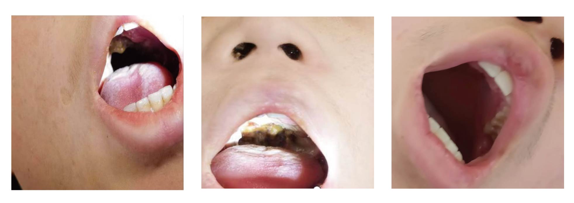
Figure 4. Significant reduction in tumor lesions observed via MRI before and one month after treatment (autologous hematopoietic stem cell transplantation combined with chemotherapy).
Discussion
ENKTCL-NT is a subtype of non-Hodgkin lymphoma and constitutes a specific form of peripheral T-cell lymphoma [1]. Lesions may involve the skin, oral cavity, gastrointestinal tract, and testes; however, rare cases have also been reported in atypical sites such as the eyes and heart. Patients presenting with ocular symptoms, including diplopia or visual disturbances, or with cardiac manifestations, such as chest tightness, pose significant diagnostic challenges. A definitive diagnosis often necessitates neuro-ophthalmological assessment, biopsy, and cardiac puncture [2]. The etiology of this disease is strongly associated with EBV infection. Accumulating evidence suggests that plasma EBV DNA load may serve as a valuable prognostic biomarker for ENKTCL-NT. Furthermore, clinical studies have established that ENKTCL-NT represents a highly aggressive malignancy with a well-documented pathogenic link to EBV infection The etiology of this disease is closely linked to EBV infection[3]. ENKTCL-NT remains a relatively rare lymphoma, with a higher incidence in East Asia and specific areas of Latin America. Evidence also points to a genetic predisposition to ENKTCL-NT, with notable ethnic and geographical patterns [4]. Consequently, diagnosing this condition in nonendemic areas poses significant challenges, often leading to underdiagnosis or misdiagnosis.
In this case, the tumor originated in the nasal cavity and subsequently invaded the oral cavity and upper jaw. Owing to the lack of noticeable symptoms in the early stages, the condition was initially overlooked. The patient sought medical attention at several hospitals and was initially misdiagnosed with sinusitis and other conditions. As the disease progressed, the patient developed oral and nasal bleeding, ultimately resulting in hemorrhagic shock due to massive hemorrhage. Early characteristic imaging findings of ENKTCL-NT, can facilitate the timely detection of the disease, underscoring the importance of identifying these radiological features. Studies indicate that about 25% of patients are accurately diagnosed with lymphoma on initial CT, around 50% are misdiagnosed, and the remaining 25% receive an indeterminate diagnosis [5]. PET-CT plays a vital role not only in the early identification of ENKTCL-NT but also in prognostic assessment. PET-CT demonstrated elevated FDG metabolism in the cervical lymph nodes, which prompted further biopsy and enabled a definitive diagnosis.
HPS is a rare disorder marked by an intense systemic inflammatory response resulting from persistent and ineffective immune activation, with hemophagocytosis serving as its defining characteristic. Common subtypes of HPS include localized aggressive hematological syndrome (LAHS), rheumatologic disease-related HPS, and EBV-associated HPS [6]. LAHS represents a highly aggressive and life-threatening clinical condition, characterized by rapid progression and a high likelihood of recurrence. The occurrence of ENKTCL-NT, in combination with HPS is extremely rare, and currently, no standardized diagnostic or therapeutic guidelines exist for this condition either nationally or internationally. Importantly, LAHS is associated with considerable mortality risk; a study involving 117 patients reported a median survival time of only 57 days. Additionally, the T/NK-LAHS subgroup exhibited a shorter median survival time compared to the B-LAHS subgroup, with survival durations of 52 days and 154 days, respectively [7]. Comprehensive treatment strategies incorporating radiotherapy have shown encouraging efficacy. Advanced radiotherapy techniques, such as volume-modulated arc therapy, provide both high therapeutic effectiveness and safety [8]. One case of splenic lymphoma-related HPS was treated using a comprehensive approach that involved a complex splenectomy in combination with chemotherapy, in the context of LAHS. The patient attained favorable clinical outcomes through timely splenectomy, followed by chemotherapy and steroid pulse therapy [9]. ASCT is frequently utilized as a subsequent therapeutic option following LAHS in combination with chemotherapy. Upon achieving disease remission, stem cell transplantation supports immune reconstitution and promotes hematopoietic recovery. ASCT is effective for both newly diagnosed and relapsed/refractory peripheral NK/T-cell lymphomas. Patients with ENKTCL-NT, particularly those with advanced-stage disease, generally face a poor prognosis.
Studies have shown that about 50% of newly diagnosed patients experience continued disease progression. Current research primarily focuses on identifying effective strategies for managing this condition and reducing relapse rates. First-line treatment combined with ASCT as consolidation therapy has been demonstrated to improve treatment efficacy and lower relapse rates. Moreover, L-asparaginase-based chemotherapy bridging ASCT has proven effective in managing ENKTCL-NT, especially in relapsed patients. In the treatment of lethal acute hemophagocytic syndrome, the timely administration of etoposide-based regimens to control hemophagocytosis, followed by ASCT upon achieving CR after chemotherapy, significantly extends progression-free survival [10]. In the present clinical case, a patient with ENKTCL-NT complicated by HPS exhibited a severe condition with rapidly progressing disease. After receiving combination chemotherapy and ASCT consolidation therapy, the patient achieved CR and remained free from recurrence during follow-up. Additionally, the patient showed favorable recovery following oral-nasal cavity and frontal facial reconstruction, underscoring a treatment approach that merits broader application.
Abbreviations
ENKTCL-NT:Extranodal NK/T cell lymphoma, nasal type; HPS:hemophagocytic syndrome; ASCT:autologous hematopoietic stem cell transplantation; PET-CT:Positron emission tomography-computed tomography; FDG:fluorodeoxyglucose; CR:complete remission; EBV:Epstein-Barr virus; VMAT:volume-modulated arc therapy; LAHS:localized aggressive hematological syndrome;
Declarations
Author contributions
Mingquan Xing(First Author): writing original draft, prepare, create, or express the content for publication, especially in writing the initial draft, including substantive translation. Chunbo Feng(CO-First Author):writing review and editing, prepare, create, or express the content for publication. Weixia Wu:Provide and verify imaging data.Help with literature search.English polishing. Yanrong Zhang:Help with literature search.Revise the article. Siqi Zhang: writing review and revise the article. Xiaoxing Sun(CO-corresponding Author): supervision, supervise and revise the article Hongfeng Ge(Corresponding Author):supervision, supervise and lead the planning and execution of research activities. All authors read and approved the final manuscript.
Acknowledgements
Thank you for the collaboration of our team and the support of the fund.
Funding information
Funding: This work was supported by 1.Scientific Research Foundation of Anhui Medical University, No.2023XKJ090;2.Key research and development project of Bozhou Science and Technology Bureau,No.bzzc2024011;3.Key research and development project of Bozhou Science and Technology Bureau,No.bzzc2022013.
Ethics approval and consent to participate
Bozhou Medical Ethics Review 2022 No. 25. The patient and his father provided written informed consent
Competing Interests
The authors declare that they have no existing or potential commercial or financial relationships that could create a conflict of interest at the time of conducting this study.
Data availability
Not Applicable.
References
 Figures
 References
 Peer
Peer Information
Figure 1. CT of the patient showing thickening of the soft tissue in the right posterior wall of the nasopharynx, mild narrowing of the right parapharyngeal space, and thickening of the soft tissue in the left palate with heterogeneous density. No significant enlargement of the lymph nodes adjacent to the bilateral carotid sheaths was observed. The paranasal sinus mucosa is thickened, and flaky soft tissue density shadows are evident.
Figure 2.Pathological examination of the patient's left mandible revealed the absence of tumor cells.
Figure 3. Prior to treatment, the patient's ulcerated maxilla exhibited communication with the nasal cavity, accompanied by a significant amount of yellow secretions and pus (as shown in the first two images). The third image illustrates the condition post-treatment.
Figure 4. Significant reduction in tumor lesions observed via MRI before and one month after treatment (autologous hematopoietic stem cell transplantation combined with chemotherapy).
Peer-review Terminology
Identity transparency: Single anonymized
Reviewer interacts with: Editor
Details
This is an open access article under the terms of the Creative Commons Attribution License(http://creativecommons.org/licenses/by/4.0/), which permits use, distribution and reproduction in any medium, provided the original work is properly cited.
Publication History
Received 2025-06-19
Accepted 2025-07-30
Published 2025-08-10