



Abstract
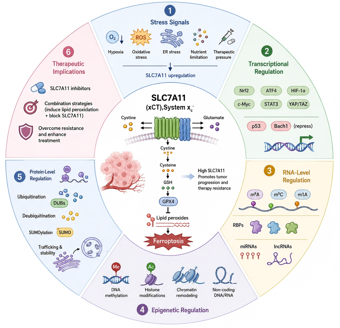
SLC7A11, also known as xCT, is the functional light-chain subunit of system xc− and mediates extracellular cystine uptake in exchange for intracellular glutamate. By supplying cystine for glutathione biosynthesis and sustaining GPX4-dependent detoxification of lipid peroxides, SLC7A11 occupies a central position in ferroptosis control. This review summarizes its multilayered regulation in cancer and highlights therapeutic strategies that combine ferroptotic pressure with blockade of adaptive SLC7A11 upregulation or stabilization.
Keywords: SLC7A11; xCT; system xc−; ferroptosis; cystine transport; glutathione; RNA modification; ubiquitination; cancer therapy
Introduction
Ferroptosis is a regulated form of cell death driven by iron-dependent lipid peroxidation. Unlike apoptosis or necrosis, ferroptosis is defined by a specific metabolic vulnerability, namely the failure to detoxify phospholipid hydroperoxides in cellular membranes. Since its initial description by Dixon et al., ferroptosis has become a major conceptual framework for understanding tumor biology, therapy resistance, metabolic stress, and redox homeostasis [1, 2]. The importance of SLC7A11 is particularly evident in cancer. Tumor cells are frequently exposed to persistent oxidative stress, hypoxia, nutrient limitation, endoplasmic reticulum stress, and therapy-induced damage. These conditions would, in principle, render malignant cells highly vulnerable to ferroptosis. However, many cancers acquire robust antioxidant programs that enable survival under such hostile conditions, and SLC7A11 is a key determinant of this adaptive phenotype. High SLC7A11 expression has been associated with tumor growth, metastasis, stemness, radioresistance, chemoresistance, and reduced sensitivity to ferroptosis-inducing therapies [3–5]. In addition, SLC7A11 reshapes amino acid metabolism, glutamine dependence, and redox balance, thereby creating both survival advantages and exploitable metabolic liabilities [3, 5]. Conversely, tumor suppressive pathways, especially p53, can repress SLC7A11 transcription and lower the threshold for ferroptosis [6].
SLC7A11 is one of the most important determinants of this adaptive state. SLC7A11, also known as xCT, is the light-chain subunit of system xc− and usually functions as a heterodimer with the heavy-chain subunit SLC3A2 [1, 2]. This transporter imports extracellular cystine into the cell in exchange for intracellular glutamate. After entering the reducing intracellular environment, cystine is converted to cysteine, which is then used for glutathione (GSH) synthesis. GSH provides reducing equivalents for glutathione peroxidase 4 (GPX4), the key enzyme that detoxifies lipid peroxides and suppresses ferroptosis [3, 7, 8]. Thus, the SLC7A11-cystine-GSH-GPX4 axis forms a core metabolic defense system against lipid peroxidation.
Accumulating evidence indicates that aberrant SLC7A11 expression in cancer is not governed by a single regulatory pathway. Rather, it is sustained by a multilayered regulatory network involving epigenetic remodeling, stress-responsive transcription factors, RNA modifications, RNA-binding proteins, non-coding RNAs, ubiquitination, deubiquitination, SUMOylation, and membrane trafficking [9–22]. Accordingly, understanding how SLC7A11 expression is regulated has become increasingly important, not only for clarifying the molecular basis of tumor stress adaptation, but also for identifying therapeutic vulnerabilities that may be exploited to sensitize tumors to ferroptosis-based treatment.
This review focuses on the expression regulation of SLC7A11 in cancer. Rather than listing individual regulators in isolation, we organize current evidence into four mechanistic layers: chromatin and epigenetic regulation, transcriptional control, RNA-level regulation, and protein-level regulation. We emphasize that SLC7A11 functions as a stress-adaptive hub through which cancer cells convert adverse microenvironmental and therapeutic signals into ferroptosis resistance. This framework also highlights a therapeutic principle: simply increasing oxidative stress may be insufficient if tumor cells simultaneously activate SLC7A11-dependent defenses. More effective strategies may require inducing ferroptotic pressure while blocking adaptive SLC7A11 upregulation or stabilization. This review is a narrative review that aims to synthesize and discuss the multilayered regulation of SLC7A11 in cancer, with a focus on its role in ferroptosis adaptation. As such, it does not follow a systematic search strategy or predefined inclusion criteria.
Structural and Functional Basis of SLC7A11 and Its Role in Tumorigenesis
SLC7A11 is the substrate-selective light-chain component of system xc− and typically forms a disulfide-linked heterodimer with the heavy-chain subunit SLC3A2 [1, 2]. Within this transporter complex, SLC3A2 mainly contributes to membrane trafficking and structural stabilization, whereas SLC7A11 confers the transport specificity required for cystine/glutamate exchange [1, 2]. SLC7A11 is a 12-transmembrane-domain protein with both its N- and C-terminal domains localized to the cytoplasm, whereas SLC3A2 is a single-pass transmembrane protein with an intracellular N-terminus and a heavily glycosylated extracellular C-terminus [23]. This structural arrangement underlies the functional output of system xc− at the plasma membrane.
SLC7A11 is positioned upstream of one of the most important anti-ferroptotic metabolic axes in cancer cells. By mediating cystine uptake, SLC7A11 ensures the intracellular supply of cysteine, a rate-limiting precursor for GSH biosynthesis. GSH then supports GPX4-dependent reduction of lipid hydroperoxides, thereby preventing lethal membrane damage. Inhibition of system xc− by genetic depletion of SLC7A11 or pharmacological blockade reduces cystine availability, decreases GSH synthesis, impairs GPX4 activity, and drives the accumulation of lipid peroxides [3, 7, 8].
This metabolic role explains why SLC7A11 is frequently associated with therapy resistance. Many anticancer treatments increase reactive oxygen species, perturb mitochondrial metabolism, or induce lipid peroxidation. Tumor cells with high SLC7A11 expression are better equipped to buffer these pressures. In this setting, SLC7A11 acts as a key buffer, allowing cancer cells to sustain redox homeostasis despite ongoing oxidative injury [3–5]. However, this protection comes with trade-offs. Because system xc− exports glutamate while importing cystine, strong SLC7A11 activity can influence glutamate metabolism and create specific nutrient dependencies. These metabolic liabilities may be exploited therapeutically, particularly in tumors that are highly dependent on cystine uptake and GSH synthesis.
SLC7A11 also functions as a readout of stress-adaptive transcriptional activity. Hypoxia, oxidative stress, endoplasmic reticulum stress, nutrient stress, inflammatory cues, and oncogenic signaling can all converge on SLC7A11 induction [9–16, 24–30]. The resulting increase in cystine uptake allows tumor cells to transform potentially damaging stress signals into antioxidant capacity. In this sense, high SLC7A11 expression should not be interpreted only as a static biomarker. It represents a dynamic adaptive state maintained by coordinated transcriptional, post-transcriptional, and post-translational regulation.
Epigenetic Regulation of SLC7A11: Chromatin Remodeling and Histone Modifications
In addition to being regulated by stress-responsive transcription factors, the expression of SLC7A11 is subject to epigenetic control at the level of chromatin architecture and histone modification. These mechanisms shape the accessibility of the SLC7A11 locus and the permissiveness of its transcriptional activation without altering the underlying DNA sequence.
Transcription of SLC7A11 depends on a permissive chromatin environment. ATP-dependent chromatin remodeling is an important component of this process. In addition to binding the promoter, NRF2 can recruit SNF2L, the ATPase subunit of the ISWI chromatin remodeling complex, to the SLC7A11 locus [31]. SNF2L promotes nucleosome repositioning and increases local chromatin accessibility, thereby facilitating transcriptional activation. This finding suggests that NRF2 functions not only as a transcription factor but also as a coordinator of chromatin remodeling.
The SWI/SNF complex also contributes to SLC7A11 regulation. Its subunit ARID1A binds the SLC7A11 promoter and supports transcriptional activation. Notably, although ARID1A is widely regarded as a tumor suppressor and is frequently mutated in cancer, its loss reduces rather than increases SLC7A11 expression, in contrast to the effects of p53 or BAP1 deficiency [32]. This highlights the context dependence of epigenetic regulators. Because ARID1A-deficient cells exhibit reduced SLC7A11 expression and impaired glutathione metabolism, they may be particularly vulnerable to therapies targeting redox homeostasis [32].
Histone modifications provide an additional layer of control. In breast cancer, HNF4α recruits p300/CBP to the SLC7A11 promoter, promotes histone acetylation, and activates SLC7A11 transcription, thereby suppressing ferroptosis [33]. By contrast, the repressive mark H3K27me3 inhibits SLC7A11 expression. A recent study showed that m6A-dependent regulation of KDM6B and GATA3 reduces KDM6B-mediated H3K27me3 demethylation and weakens GATA3-driven transactivation, resulting in coordinated suppression of SLC7A11 [34]. This mechanism illustrates how RNA modification, histone regulation, and transcription factor activity can converge on the same locus.
Overall, chromatin remodeling and histone modification collectively determine the accessibility and transcriptional permissiveness of the SLC7A11 locus, and thereby set the baseline upon which stress-responsive transcription factors act.
Transcriptional Regulatory Network of SLC7A11
Transcriptional control constitutes the most direct mechanism by which cancer cells increase SLC7A11 expression in response to environmental and therapeutic stress. Multiple signaling pathways converge on the SLC7A11 locus, forming an integrated regulatory network that determines ferroptosis sensitivity.
KEAP1–NRF2 signaling as a central antioxidant axis
Among all transcriptional regulators, the KEAP1–NRF2 pathway is one of the most prominent drivers of SLC7A11 expression. Under basal conditions, KEAP1 promotes NRF2 degradation, whereas oxidative stress or KEAP1 dysfunction stabilizes NRF2 and enables its nuclear translocation. Activated NRF2 binds antioxidant response elements and induces expression of a wide range of cytoprotective genes, including SLC7A11 [24]. In many cancers, persistent NRF2 activation enhances cystine uptake, promotes GSH production, and suppresses ferroptosis, thereby facilitating tumor survival under oxidative stress.
Recent studies further indicate that the NRF2–SLC7A11 axis is shaped by metabolic and microenvironmental cues. In triple-negative breast cancer, cysteine availability itself can promote NRF2 activation, forming a feed-forward survival mechanism [25]. In gastric cancer, TMEM160 suppresses ferroptosis and induces chemoresistance by inhibiting KEAP1 and thereby activating NRF2-dependent SLC7A11 expression [26]. Lipid metabolism also intersects with this pathway, as SCD1 inhibition in lung adenocarcinoma blocks AKT–NRF2–SLC7A11 signaling and primes cells for ferroptosis [27]. Moreover, immune-metabolic signals from the tumor microenvironment can enhance ferroptosis resistance through SLC7A11-associated antioxidant programs [28]. These findings suggest that NRF2-dependent induction of SLC7A11 is not a simple generic oxidative stress response, but rather part of a broader adaptive network integrating metabolism, redox regulation, and therapy pressure [24–30] (Figure 1A).
Hypoxia, HIF signaling, and ATF4-Mediated integrated stress response
Hypoxia is a hallmark of solid tumors and an important driver of SLC7A11 transcription. HIF-1 can promote glutathione synthesis and ferroptosis resistance by inducing SLC7A11-associated transcriptional programs, thereby contributing to therapy-resistant cancer stem-like phenotypes [9]. HIF-2 may also mediate ferroptosis resistance in a tumor type-specific manner, as reported in pancreatic cancer [10]. Although the relative contribution of HIF-1 versus HIF-2 may vary across contexts, these studies support the general concept that hypoxia-induced SLC7A11 expression is an important component of tumor adaptation.
HIF signaling also cooperates with oncogenic metabolic pathways. In ovarian cancer, YY1-induced USP43 stabilizes FASN, which subsequently activates SLC7A11 expression through HIF-1α stabilization [11]. This cascade illustrates how lipid metabolism and deubiquitinating pathways can converge on hypoxia-responsive transcriptional control. Thus, SLC7A11 upregulation under hypoxia reflects an active integration of oxygen sensing, transcription factor stability, and metabolic rewiring rather than a passive consequence of low oxygen tension.
Endoplasmic reticulum stress and nutrient limitation provide an additional route to SLC7A11 induction via ATF4, a central transcription factor of the integrated stress response. ATF4 directly activates SLC7A11 transcription in hepatocellular carcinoma and colorectal cancer, thereby promoting cystine uptake and GSH synthesis under adverse conditions [12, 13]. Pharmacological evidence further supports this pathway, as dihydroartemisinin induces ferroptosis in hepatocellular carcinoma partly by suppressing the ATF4–xCT axis [14]. Given that hypoxia can simultaneously elevate reactive oxygen species and trigger endoplasmic reticulum stress, HIF and ATF4 pathways may cooperate to establish a robust stress-adaptive program sustaining SLC7A11 expression in solid tumors (Figure 1B).
p53 as a context-dependent transcriptional brake
In contrast to the activating pathways described above, p53 serves as a major transcriptional repressor of SLC7A11. p53 directly binds the SLC7A11 promoter and represses its transcription, thereby reducing cystine uptake, limiting GSH synthesis, and sensitizing cells to ferroptosis [6]. This mechanism links tumor suppressor activity to ferroptosis regulation and is distinct from the classical roles of p53 in cell-cycle arrest, apoptosis, and senescence.
Importantly, the p53–SLC7A11 axis is highly context dependent. Its output may be influenced by p53 mutation status, polymorphisms, post-translational modifications, interacting cofactors, and tumor-specific metabolic context. For example, the African-specific P47S variant of p53 exhibits impaired tumor suppressor function and altered ferroptosis regulation [17]. ACSL1 loss in clear cell renal cell carcinoma modifies ferroptosis resistance in a manner connected to lipid metabolism [18]. In ovarian cancer, TOP2A knockdown reverses cisplatin resistance through the TP53/GPX4/SLC7A11 axis, whereas in breast cancer FXR deficiency induces ferroptosis by modulating CBP-dependent p53 acetylation [19, 20]. Additional regulators such as CAPG and lncRNA-HMG further reshape p53-mediated ferroptosis through broader signaling and non-coding RNA networks [21, 22]. These findings indicate that p53 is best viewed as a context-sensitive ferroptosis gatekeeper rather than a universal binary switch (Figure 1C).
Additional transcriptional inputs
In addition to canonical stress pathways, several other mechanisms contribute to SLC7A11 transcriptional activation. Under oxidative stress, ROS can trigger an oxidation–O-GlcNAcylation cascade that promotes FOXK2 nuclear translocation and SLC7A11 transcription, representing an NRF2-independent route by which oxidative injury is converted into antioxidant adaptation [15]. In estrogen receptor-positive breast cancer, estrogen receptor α enhances system xc− activity and SLC7A11 expression, thereby linking hormone signaling to ferroptosis resistance and endocrine responsiveness [16]. These findings broaden the conceptual framework of SLC7A11 regulation, indicating that distinct tumor types may use different upstream routes to converge on the same anti-ferroptotic effector.
Taken together, the transcriptional regulatory network of SLC7A11 integrates oxidative stress, hypoxia, endoplasmic reticulum stress, nutrient availability, hormonal cues, and tumor suppressor pathways to determine whether tumor cells remain resistant or become vulnerable to ferroptosis (Figure 1D).
Figure 1. Transcriptional regulation of SLC7A11.
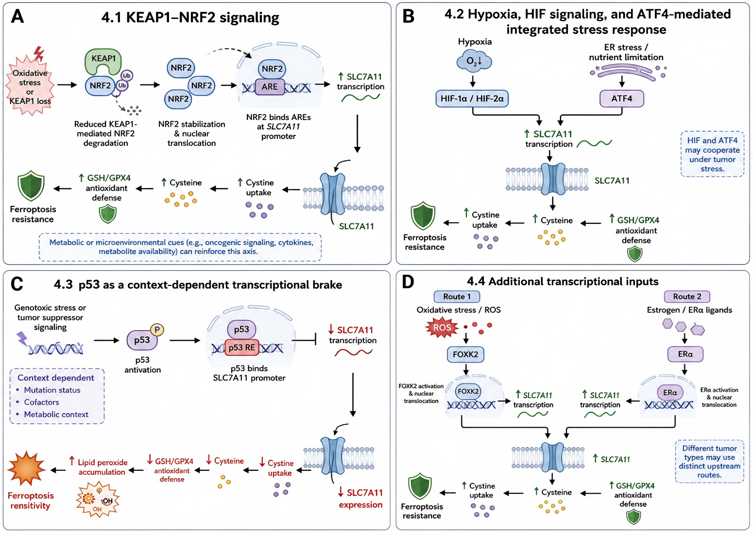
A, KEAP1 loss or oxidative stress stabilizes NRF2, promoting SLC7A11 transcription and ferroptosis resistance. B, hypoxia, ER stress and nutrient limitation activate HIFs or ATF4 to induce SLC7A11 expression. C, p53 represses SLC7A11 transcription, reducing cystine uptake and increasing ferroptosis sensitivity. D, ROS–FOXK2 and estrogen–ERα signaling provide additional routes for SLC7A11 activation.
Post-Transcriptional and Translational Regulation of SLC7A11
After transcription, SLC7A11 expression is further refined at the RNA level through RNA modifications, RNA-binding proteins, non-coding RNAs, and alternative splicing. This layer of regulation allows cells to rapidly and reversibly adjust SLC7A11 output in response to environmental fluctuation and therapeutic challenge.
RNA modifications and mRNA fate
Among epitranscriptomic modifications, m6A is the most extensively studied in the context of SLC7A11. METTL3-mediated m6A modification of SLC7A11 enhances radioresistance in nasopharyngeal carcinoma by inhibiting ferroptosis [35]. In non-small cell lung cancer, IGF2BP2 recognizes m6A-modified SLC7A11 mRNA and promotes its stability, thereby supporting proliferation and ferroptosis resistance [36]. METTL5 likewise promotes cervical cancer progression through m6A-dependent suppression of ferroptosis involving SLC7A11 [37]. In hepatocellular carcinoma, inhibition of METTL3 enhances radiosensitivity by downregulating SLC7A11, while IGF2BP3 stabilizes SLC7A11 in gastric cancer and antagonizes ferroptosis [38, 39].
Conversely, RNA demethylases can reduce SLC7A11 expression and increase ferroptosis sensitivity. ALKBH5 suppresses non-small cell lung cancer progression by facilitating ferroptosis through m6A demethylation of SLC7A11 [40]. FTO inhibition decreases the SLC7A11/GPX4 axis and induces ferroptotic death in colorectal cancer [41]. These observations emphasize that the effect of m6A on SLC7A11 is highly context dependent and may vary according to the modified site, reader proteins, tumor lineage, and surrounding stress environment.
Other RNA modifications also participate in SLC7A11 control. NSUN2-mediated m5C modification affects SLC7A11 mRNA stability and translation, and NSUN2 knockdown promotes ferroptosis in colorectal cancer [42]. NAT10-mediated ac4C modification increases SLC7A11 protein expression and contributes to sorafenib resistance in nasopharyngeal carcinoma [43]. Together, these studies identify SLC7A11 as an important effector transcript in the broader field of epitranscriptomic regulation of ferroptosis (Figure 2A).
RNA-binding proteins and 3′UTR-mediated regulation
The stability and translational efficiency of SLC7A11 mRNA are also strongly influenced by RNA-binding proteins interacting with cis-regulatory elements, particularly within the 3′ untranslated region (3′UTR). In bladder cancer, PHGDH promotes malignant progression by upregulating SLC7A11, at least partly through stabilization of the RNA-binding protein PCBP2 [44]. In colon adenocarcinoma, HnRNPU stabilizes the mRNAs encoding both SLC7A11 and SLC3A2, suggesting coordinated control of the two subunits of system xc− [45]. ALYREF similarly promotes SLC7A11 mRNA stability and facilitates lung adenocarcinoma growth and metastasis [46].
By contrast, destabilizing RNA-binding proteins can decrease SLC7A11 levels and sensitize cells to ferroptosis. In KRASG12C-mutant lung cancer, TTP binds AU-rich elements in the SLC7A11 3′UTR and accelerates mRNA decay, thereby enhancing sensitivity to KRASG12C inhibitors [47]. RBMS2 has also been reported to regulate SLC7A11 transcription–translation coupling in colorectal cancer [48]. These studies underscore that the 3′UTR of SLC7A11 functions as a regulatory platform where oncogenic signaling, metabolic status, and therapy response are integrated (Figure 2B).
Non-coding RNAs and alternative splicing
Non-coding RNAs constitute another major post-transcriptional regulatory layer upstream of SLC7A11. Many long non-coding RNAs (lncRNAs) and circular RNAs (circRNAs) act through competitive endogenous RNA mechanisms, typically by sequestering microRNAs that would otherwise repress SLC7A11. In colorectal cancer, lncRNA FTX promotes radioresistance through the miR-625-5p/SLC7A11 axis [49]. In lung cancer, small extracellular vesicle-associated SNHG12 enhances metastasis through the miR-326/SLC7A11 axis, and lncRNA SLC7A11AR promotes lung adenocarcinoma progression by increasing SLC7A11 expression [50, 51]. In breast cancer, POU2F2-induced PTPRG-AS1 inhibits ferroptosis through the miR-376c-3p/SLC7A11 axis [52]. In hepatocellular carcinoma, circTTC13 promotes sorafenib resistance through the miR-513a-5p/SLC7A11 axis [53].
Notably, non-coding RNAs can also regulate SLC7A11 through mechanisms beyond classical miRNA sponging. LINC01833 protects SLC7A11 from WWP1-mediated ubiquitination and promotes gemcitabine resistance in non-small cell lung cancer [54]. LINC01088 enhances SLC7A11 expression in glioblastoma through the HLTF/USP7 axis [55]. Under glutamine starvation, the lncRNA FERRIN suppresses ferroptosis by stabilizing SLC7A11 mRNA [56]. Exosome-derived circUPF2 enhances resistance to targeted therapy in hepatocellular carcinoma by strengthening the interaction between IGF2BP2 and SLC7A11 mRNA [57]. In prostate cancer, circCNOT6L modulates alternative splicing of SLC7A11 pre-mRNA through SRSF2, thereby extending post-transcriptional regulation from mRNA abundance to transcript processing [58].
Collectively, these findings indicate that non-coding RNAs influence SLC7A11 at multiple levels, including transcript stability, protein turnover, and RNA processing, and that post-transcriptional and translational regulation provides a rapid and versatile means for cancer cells to fine-tune SLC7A11 expression and dynamically adjust ferroptosis sensitivity. (Figure 2C).
Figure 2. Post-transcriptional and translational regulation of SLC7A11.
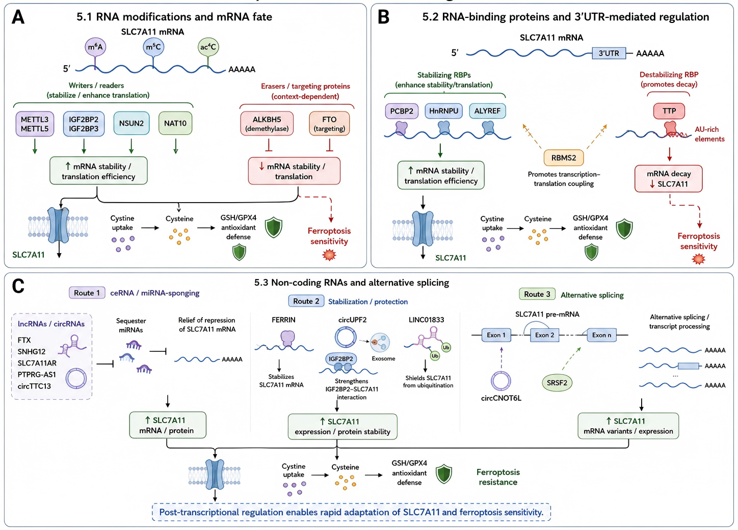
A, RNA modifications regulate SLC7A11 mRNA stability and translation, thereby altering ferroptosis sensitivity. B, RNA-binding proteins control SLC7A11 mRNA stability through 3′UTR-dependent mechanisms. C, non-coding RNAs and alternative splicing modulate SLC7A11 expression, protein stability and ferroptosis resistance.
Protein Homeostasis and Post-Translational Regulation of SLC7A11
Although transcriptional and RNA-level mechanisms determine SLC7A11 mRNA output, the ultimate anti-ferroptotic function of SLC7A11 depends on protein abundance, stability, membrane localization, and transporter complex assembly.
Ubiquitination-mediated degradation
The ubiquitin–proteasome system is a major mechanism governing SLC7A11 protein turnover. Several E3 ubiquitin ligases promote SLC7A11 degradation and thereby enhance ferroptosis sensitivity. In hepatocellular carcinoma, SOCS2 induces K48-linked polyubiquitination of SLC7A11 by recruiting the Elongin B/C complex, leading to its degradation and consequent radiosensitization [59]. In colorectal cancer, AMER1 recruits β-TrCP1/2 to target SLC7A11 and FTL, thereby promoting ferroptosis and suppressing metastasis [60]. TRIM3 facilitates K11-linked ubiquitination and degradation of SLC7A11 in non-small cell lung cancer [61]. ZRANB1 has also been reported to act as an E3 ligase for SLC7A11 and to regulate ferroptotic resistance [62]. These studies indicate that distinct tumors may employ different ubiquitin ligases and linkage types to regulate SLC7A11 stability (Figure 3A).
Deubiquitination, SUMOylation, and protein stabilization
Deubiquitinating enzymes (DUBs) can counteract ubiquitin-mediated degradation and maintain SLC7A11 abundance. OTUD5 removes ubiquitin chains from SLC7A11 in triple-negative breast cancer, promoting progression and reducing paclitaxel sensitivity [63]. OTUD3 stabilizes SLC7A11 and contributes to sunitinib resistance in clear cell renal cell carcinoma [64]. In multiple myeloma, HSPA9 enhances USP14-mediated SLC7A11 deubiquitination and suppresses ferroptosis [65]. In hepatocellular carcinoma, EFNA4 recruits USP9X to stabilize SLC7A11, linking extracellular signaling to intracellular ferroptosis resistance [66]. USP35 promotes growth of estrogen receptor-positive breast cancer through a BRD4-SLC7A11-dependent anti-ferroptotic mechanism [67] (Figure 3B).
SUMOylation is another important post-translational mechanism. PIAS4-mediated SUMOylation stabilizes SLC7A11 in breast cancer, whereas tanshinone IIA destabilizes SLC7A11 by interfering with KDM1A–PIAS4-dependent SUMOylation and thereby promotes ferroptosis [68]. These results suggest that protein stabilization pathways may sustain SLC7A11 expression even when transcriptional activation is only moderate, making them attractive therapeutic targets.
Membrane localization and system xc− complex activity
The functional output of SLC7A11 depends not only on total protein abundance but also on its localization to the plasma membrane and its proper assembly with SLC3A2 [1, 2]. SLC3A2 is required for transporter stability and trafficking, whereas SLC7A11 provides the substrate-selective transport function. Coordinated regulation of both subunits may therefore be necessary for maximal cystine uptake. For example, HnRNPU stabilizes both SLC7A11 and SLC3A2 mRNAs, supporting the idea that the entire system xc− complex can be co-regulated [45] (Figure 3C).
Compared with transcriptional, RNA-level, and ubiquitin-dependent regulation, mechanisms governing SLC7A11 trafficking, membrane retention, internalization, recycling, and lysosomal degradation remain less clearly defined. This represents an important knowledge gap, because only the membrane-localized fraction of SLC7A11 is functionally active. Future studies integrating surface biotinylation, membrane proteomics, live-cell imaging, and cystine uptake assays will be needed to distinguish total SLC7A11 protein from surface-functional SLC7A11 under stress and treatment conditions (Figure 3D). Representative regulators across different regulatory layers of SLC7A11 are summarized in Table 1.
Figure 3. Protein-level regulation of SLC7A11.
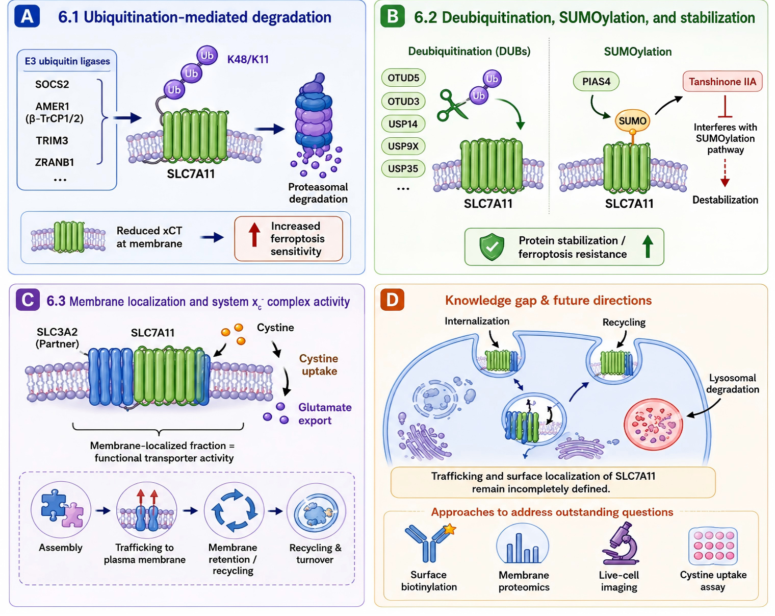
A, E3 ligases mediate SLC7A11 ubiquitination and degradation, reducing membrane xCT and increasing ferroptosis sensitivity. B, DUBs and SUMOylation stabilize SLC7A11, promoting ferroptosis resistance. C, SLC3A2-assisted membrane localization enables system xc⁻ activity, cystine uptake and glutamate export. D, SLC7A11 trafficking, recycling and lysosomal degradation remain key unresolved mechanisms for future study.
Therapeutic implications: Inducing Ferroptotic Pressure while Blocking Adaptive Defense
The regulatory network described above has an important therapeutic implication: ferroptosis-inducing therapy may fail if tumor cells simultaneously activate SLC7A11-dependent adaptive defenses. Many treatments increase oxidative stress, hypoxia, lipid peroxidation, endoplasmic reticulum stress, or metabolic pressure. These same stimuli can activate NRF2, HIF, ATF4 [9–14, 24–27], RNA stabilization programs [35–48], and protein stabilization mechanisms [59–68] that restore or further increase SLC7A11 expression. As a result, treatment-induced stress may paradoxically select for SLC7A11-high, ferroptosis-resistant cancer cell populations [3–5, 24].
A more effective therapeutic principle may be to combine stress induction with adaptation blockade. The first component increases lipid peroxidation pressure through radiotherapy, chemotherapy, targeted therapy, GPX4 inhibition, system xc− inhibition, or agents that perturb lipid metabolism. The second component blocks the compensatory SLC7A11 response at one or more regulatory levels. Potential targets include NRF2 or KEAP1-dependent transcriptional activation, HIF-driven hypoxia adaptation, ATF4-mediated integrated stress response, m6A writer-reader modules, stabilizing RNA-binding proteins, oncogenic non-coding RNAs, DUBs, and SUMOylation enzymes. Regarding the first component, several clinically available agents can, in principle, serve as the ferroptosis-inducing arm of such combination regimens. Sorafenib, a multikinase inhibitor approved for hepatocellular carcinoma, renal cell carcinoma, and thyroid cancer, induces ferroptosis partly through system xc- suppression and GSH depletion [69]. By contrast, therapeutic strategies targeting the second component—namely, blockade of adaptive SLC7A11 upregulation or stabilization—currently lack clinical-stage compounds and remain largely at the preclinical or target-validation stage. Bridging this gap will require not only the development of selective and pharmacologically tractable inhibitors, but also biomarker-driven patient selection strategies to identify tumors in which SLC7A11-dependent adaptive defense is the dominant resistance mechanism. Whether such combination approaches are ultimately feasible will need to be validated in further preclinical and clinical studies.
This strategy also requires biomarker development. SLC7A11 expression alone may be insufficient to stratify patients because its activity is shaped by upstream drivers and post-transcriptional regulators. A more informative biomarker panel may include SLC7A11 protein level, membrane localization, SLC3A2 co-expression, KEAP1/NRF2 status, p53 status, ATF4/HIF activity, GSH abundance, lipid peroxide burden, and expression of key RNA or protein stabilizers. Such integrated profiling could help identify tumors that are both dependent on SLC7A11 and vulnerable to combined ferroptosis-based interventions.
Several challenges remain. First, SLC7A11 regulation is highly context dependent [3, 6, 17]. The same regulator may have different effects depending on tumor lineage, genetic background, nutrient availability, and treatment exposure. Second, systemic inhibition of cystine uptake or antioxidant defense may cause toxicity in normal tissues [2, 3]. Third, ferroptosis does not occur in isolation. It interacts with apoptosis, immune signaling, metabolic remodeling, and the tumor microenvironment [7, 8, 28]. Therefore, future therapeutic studies should incorporate in vivo models, patient-derived organoids, immune-competent systems, single-cell omics, and spatial analysis to determine when SLC7A11-targeted strategies are most likely to succeed.
Conclusions and future perspectives
SLC7A11 is a central regulator of ferroptosis sensitivity in cancer. By importing extracellular cystine and supporting GSH-GPX4-dependent lipid peroxide detoxification, it allows tumor cells to maintain redox homeostasis under hostile microenvironmental and therapeutic conditions. Current evidence indicates that high SLC7A11 expression is not produced by a single pathway. It is maintained by coordinated regulation at the transcriptional, chromatin, RNA, and protein levels.
At the transcriptional level, NRF2, HIFs, ATF4, hormone receptors, FOXK2, and oncogenic metabolic pathways activate SLC7A11 in response to oxidative stress, hypoxia, endoplasmic reticulum stress, and metabolic remodeling. In contrast, p53 functions as a context-dependent transcriptional brake that links tumor suppression to ferroptosis sensitivity. At the chromatin level, remodeling factors, histone acetylation programs, and chromatin-associated transcriptional coactivators shape accessibility of the SLC7A11 locus. At the RNA level, m6A, m5C, ac4C, RNA-binding proteins, non-coding RNAs, and splicing regulators determine mRNA stability, translation, and isoform output. At the protein level, ubiquitination, deubiquitination, SUMOylation, and membrane localization determine whether SLC7A11 becomes a functional transporter at the cell surface. Notably, total SLC7A11 expression does not necessarily reflect membrane-localized transporter activity, and the mechanisms governing this gap remain an important open question for future investigation.
The central message is that SLC7A11 acts as a stress-adaptive hub. Tumor cells use this hub to transform oxidative stress, hypoxia, nutrient stress, and therapy-induced damage into antioxidant defense and ferroptosis resistance. This concept also reframes therapy. Rather than relying only on agents that increase oxidative or lipid peroxidation stress, future strategies may need to pair ferroptosis induction with blockade of SLC7A11 adaptation. Such approaches may include targeting upstream transcriptional activators, RNA-stabilizing machinery, non-coding RNA networks, DUBs, SUMOylation pathways, or transporter membrane function.
Future research should move beyond single-regulator studies and define the hierarchy, timing, and cooperativity of SLC7A11 regulatory modules in specific tumor contexts. Integrating single-cell transcriptomics, spatial proteomics, metabolomics, functional genetic screening, and patient-derived models will be essential for identifying actionable SLC7A11-high states. A deeper understanding of this multilayered regulatory architecture may provide a foundation for more precise and durable ferroptosis-targeted cancer therapies.
Table 1. Representative regulators of SLC7A11 expression and functional consequences
| Regulatory layer | Representative regulators | Mechanism and functional consequence | Key references |
|---|---|---|---|
| Transcriptional control | NRF2, HIFs, ATF4, p53, ERα, FOXK2 | Stress-responsive transcriptional activation or repression of SLC7A11; balances antioxidant adaptation and ferroptosis sensitivity. | [6, 9–16, 24–30] |
| Chromatin and epigenetic control | SNF2L, ARID1A, HNF4α-p300/CBP, KDM6B/GATA3 | Controls promoter accessibility, histone acetylation, chromatin state, and sustained transcriptional output. | [31–34] |
| RNA modifications | METTL3, METTL5, IGF2BP2/3, ALKBH5, FTO, NSUN2, NAT10 | Regulates SLC7A11 mRNA stability, translation efficiency, and therapy-associated ferroptosis resistance. | [35–43] |
| RNA-binding proteins and non-coding RNAs | PCBP2, HnRNPU, ALYREF, TTP, RBMS2, FTX, SNHG12, SLC7A11AR, circUPF2, circCNOT6L, circTTC13 | Controls mRNA decay, stabilization, miRNA-dependent repression, scaffold functions, and alternative splicing. | [44–58] |
| Protein-level regulation | SOCS2, AMER1, TRIM3, ZRANB1, OTUD5, OTUD3, USP14, USP9X, USP35, PIAS4 | Determines SLC7A11 degradation, stabilization, SUMOylation, membrane availability, and functional anti-ferroptotic output. | [59–68] |
Abbreviations
3′UTR: 3′ Untranslated Region; ac4C: N4-Acetylcytidine; ACSL1: Acyl-CoA Synthetase Long Chain Family Member 1; AKT: AKT Serine/Threonine Kinase; ALKBH5: AlkB Homolog 5; ALYREF: Aly/REF Export Factor; AMER1: APC Membrane Recruitment Protein 1; ARID1A: AT-Rich Interaction Domain 1A; ATF4: Activating Transcription Factor 4; BAP1: BRCA1 Associated Protein 1; BRD4: Bromodomain Containing 4; CAPG: Capping Actin Protein, Gelsolin Like; CBP: CREB Binding Protein; circCNOT6L: Circular RNA from CNOT6L Gene; circRNA: Circular RNA; circTTC13: Circular RNA from TTC13 Gene; circUPF2: Circular RNA from UPF2 Gene; DUB: Deubiquitinating Enzyme; ERα: Estrogen Receptor Alpha; FOXK2: Forkhead Box K2; FTL: Ferritin Light Chain; FTO: Fat Mass and Obesity-Associated Protein; FXR: Farnesoid X Receptor; GATA3: GATA Binding Protein 3; GPX4: Glutathione Peroxidase 4; GSH: Glutathione; H3K27me3: Histone H3 Lysine 27 Trimethylation; HIF: Hypoxia-Inducible Factor; HIF-1: Hypoxia-Inducible Factor 1; HIF-2: Hypoxia-Inducible Factor 2; HLTF: Helicase-Like Transcription Factor; HNF4α: Hepatocyte Nuclear Factor 4 Alpha; HnRNPU: Heterogeneous Nuclear Ribonucleoprotein U; HSPA9: Heat Shock Protein Family A Member 9; IGF2BP2: Insulin Like Growth Factor 2 mRNA Binding Protein 2; IGF2BP3: Insulin Like Growth Factor 2 mRNA Binding Protein 3; KDM6B: Lysine Demethylase 6B; KEAP1: Kelch Like ECH Associated Protein 1; LINC01088: Long Intergenic Non-Protein Coding RNA 1088; LINC01833: Long Intergenic Non-Protein Coding RNA 1833; lncRNA: Long Non-Coding RNA; m5C: 5-Methylcytosine; m6A: N6-Methyladenosine; METTL3: Methyltransferase Like 3; METTL5: Methyltransferase Like 5; miRNA: MicroRNA; NAT10: N-Acetyltransferase 10; NRF2: Nuclear Factor Erythroid 2-Related Factor 2; NSUN2: NOP2/Sun RNA Methyltransferase 2; OTUD3: Ovarian Tumor Deubiquitinase 3; OTUD5: Ovarian Tumor Deubiquitinase 5; p300: E1A Binding Protein p300; PCBP2: Poly(rC) Binding Protein 2; PHGDH: Phosphoglycerate Dehydrogenase; PIAS4: Protein Inhibitor of Activated STAT 4; POU2F2: POU Class 2 Homeobox 2; PTPRG-AS1: PTPRG Antisense RNA 1; RBMS2: RNA Binding Motif Single Stranded Interacting Protein 2; ROS: Reactive Oxygen Species; SCD1: Stearoyl-CoA Desaturase 1; SLC3A2: Solute Carrier Family 3 Member 2; SLC7A11: Solute Carrier Family 7 Member 11; SNF2L: SWI/SNF Related, Matrix Associated, Actin Dependent Regulator of Chromatin, Subfamily A-Like 1; SNHG12: Small Nucleolar RNA Host Gene 12; SOCS2: Suppressor of Cytokine Signaling 2; SRSF2: Serine and Arginine Rich Splicing Factor 2; SUMOylation: Small Ubiquitin-Like Modifier Conjugation; SWI/SNF: Switch/Sucrose Non-Fermentable; system xc−: Cystine/Glutamate Antiporter System; TMEM160: Transmembrane Protein 160; TOP2A: DNA Topoisomerase II Alpha; TP53: Tumor Protein p53; TRIM3: Tripartite Motif Containing 3; TTP: Tristetraprolin; USP14: Ubiquitin Specific Peptidase 14; USP35: Ubiquitin Specific Peptidase 35; USP43: Ubiquitin Specific Peptidase 43; USP7: Ubiquitin Specific Peptidase 7; USP9X: Ubiquitin Specific Peptidase 9 X-Linked; WDR74: WD Repeat Domain 74; WWP1: WW Domain Containing E3 Ubiquitin Protein Ligase 1; xCT: Cystine/Glutamate Antiporter; ZRANB1: Zinc Finger RANBP2-Type Containing 1; β-TrCP1/2: Beta-Transducin Repeat Containing E3 Ubiquitin Protein Ligase 1/2.
Declarations
Acknowledgements
Not Applicable.
Ethics Approval and Consent to Participate
We declare that our manuscript is not published elsewhere. All co-authors had substantial contribution to the content and creation of the manuscript. All authors approved the final version of the submitted paper.
Funding Information
Not Applicable.
Author Contributions
ZHY conceptualized, researched, wrote, and edited the entire manuscript.
Data Availability
Not Applicable.
Competing Interests
The authors declare that they have no existing or potential commercial or financial
relationships that could create a conflict of interest at the time of conducting this study.
References
[1] Sato, H, M Tamba, K Kuriyama-Matsumura, S Okuno, and S Bannai. Molecular cloning and expression of human xCT, the light chain of amino acid transport system xc-. Antioxid. Redox Signal. 2000;2(4):665–671. https://doi.org/10.1089/ars.2000.2.4-665
[2] Lewerenz, J, SJ Hewett, Y Huang, M Lambros, PW Gout, PW Kalivas, et al. The cystine/glutamate antiporter system x(c)(-) in health and disease: from molecular mechanisms to novel therapeutic opportunities. Antioxid. Redox Signal. 2013;18(5):522–555. https://doi.org/10.1089/ars.2011.4391
[3] Koppula, P, L Zhuang, and B Gan. Cystine transporter SLC7A11/xCT in cancer: ferroptosis, nutrient dependency, and cancer therapy. Protein Cell. 2021;12(8):599–620. https://doi.org/10.1007/s13238-020-00789-5
[4] Liu, J, X Xia, and P Huang. xCT: A Critical Molecule That Links Cancer Metabolism to Redox Signaling. Mol. Ther. 2020;28(11):2358–2366. https://doi.org/10.1016/j.ymthe.2020.08.021
[5] Koppula, P, Y Zhang, L Zhuang, and B Gan. Amino acid transporter SLC7A11/xCT at the crossroads of regulating redox homeostasis and nutrient dependency of cancer. Cancer Commun. 2018;38(1):12. https://doi.org/10.1186/s40880-018-0288-x
[6] Jiang, L, N Kon, T Li, S Wang, T Su, H Hibshoosh, et al. Ferroptosis as a p53-mediated activity during tumour suppression. Nature. 2015;520(7545):57–62. https://doi.org/10.1038/nature14344
[7] Dixon, SJ, KM Lemberg, MR Lamprecht, R Skouta, EM Zaitsev, CE Gleason, et al. Ferroptosis: an iron-dependent form of nonapoptotic cell death. Cell. 2012;149(5):1060–1072. https://doi.org/10.1016/j.cell.2012.03.042
[8] Stockwell, BR, JP Friedmann Angeli, H Bayir, AI Bush, M Conrad, SJ Dixon, et al. Ferroptosis: A Regulated Cell Death Nexus Linking Metabolism, Redox Biology, and Disease. Cell. 2017;171(2):273–285. https://doi.org/10.1016/j.cell.2017.09.021
[9] Lu, H, D Samanta, L Xiang, H Zhang, H Hu, I Chen, et al. Chemotherapy triggers HIF-1–dependent glutathione synthesis and copper chelation that induces the breast cancer stem cell phenotype. Proc. Natl. Acad. Sci. U. S. A. 2015;112(33):E4600–E4609. https://doi.org/10.1073/pnas.1513433112
[10] Hubbi, ME, CL Wang, Y Suhail, NL Almasri, J Xie, EE Hollander, et al. The HIF-2 Transcription Factor Mediates Resistance to Ferroptosis in Pancreatic Cancer. Mol. Cell. 2026;86(7):1260-1274.e4. https://doi.org/10.1016/j.molcel.2026.03.007
[11] Zhao, T, X Chen, J Wu, S Chen, Y Gan, C Liu, et al. YY1-induced USP43 drives ferroptosis suppression by FASN stabilization and subsequent activation of SLC7A11 in ovarian cancer. Cell Death Dis. 2025;16(1):589. https://doi.org/10.1038/s41419-025-07886-5
[12] Lin, Z, S Yang, Q Qiu, G Cui, Y Zhang, M Yao, et al. Hypoxia-induced cysteine metabolism reprogramming is crucial for the tumorigenesis of colorectal cancer. Redox Biol. 2024;75:103286. https://doi.org/10.1016/j.redox.2024.103286
[13] He, F, P Zhang, J Liu, R Wang, RJ Kaufman, BC Yaden, et al. ATF4 suppresses hepatocarcinogenesis by inducing SLC7A11 (xCT) to block stress-related ferroptosis. J. Hepatol. 2023;79(2):362–377. https://doi.org/10.1016/j.jhep.2023.03.016
[14] Ji, J, Z Cheng, J Zhang, J Wu, X Xu, C Guo, et al. Dihydroartemisinin induces ferroptosis of hepatocellular carcinoma via inhibiting ATF4‐xCT pathway. J. Cell. Mol. Med. 2024;28(8):e18335. https://doi.org/10.1111/jcmm.18335
[15] Zhang, H, J Ma, C Hou, X Luo, S Zhu, Y Peng, et al. A ROS-mediated oxidation-O-GlcNAcylation cascade governs ferroptosis. Nat. Cell Biol. 2025;27(8):1288–1300. https://doi.org/10.1038/s41556-025-01722-w
[16] Cao, J, T Zhou, T Wu, R Lin, J Huang, D Shi, et al. Targeting estrogen-regulated system xc− promotes ferroptosis and endocrine sensitivity of ER+ breast cancer. Cell Death Dis. 2025;16(1):30. https://doi.org/10.1038/s41419-025-07354-0
[17] Jennis, M, C-P Kung, S Basu, A Budina-Kolomets, JI-J Leu, S Khaku, et al. An African-specific polymorphism in the TP53 gene impairs p53 tumor suppressor function in a mouse model. Genes Dev. 2016;30(8):918–930. https://doi.org/10.1101/gad.275891.115
[18] Wang, S, Y Wang, B Liu, D Zhang, Z Zhang, H Yang, et al. Loss of ACSL1 fuels ferroptosis resistance in clear cell renal carcinoma. Cancer Biol. Ther. 26(1):2567815. https://doi.org/10.1080/15384047.2025.2567815
[19] Liu, X, J Wang, Y Liu, Y Han, Z Qin, Y Wang, et al. Knockdown of TOP2A reverses cisplatin resistance in ovarian cancer by inhibiting EMT via ferroptosis mediated by the TP53/GPX4/SLC7A11 axis. Front. Immunol. 2025;16:1675373. https://doi.org/10.3389/fimmu.2025.1675373
[20] Huang, P, H Zhao, H Dai, J Li, X Pan, W Pan, et al. FXR deficiency induced ferroptosis via modulation of the CBP-dependent p53 acetylation to suppress breast cancer growth and metastasis. Cell Death Dis. 2024;15(11):826. https://doi.org/10.1038/s41419-024-07222-3
[21] Quan, B, F Yao, W Liu, B Tang, M Li, S Lu, et al. Increased CAPG inhibits ferroptosis to drive tumor proliferation and sorafenib resistance in hepatocellular carcinoma via the WDR74-p53-SLC7A11 pathway. Int. J. Biol. Sci. 2025;21(12):5476–5495. https://doi.org/10.7150/ijbs.111419
[22] Xin, Z, C Hu, C Zhang, M Liu, J Li, X Sun, et al. LncRNA-HMG incites colorectal cancer cells to chemoresistance via repressing p53-mediated ferroptosis. Redox Biol. 2024;77:103362. https://doi.org/10.1016/j.redox.2024.103362
[23] Sato, H, M Tamba, T Ishii, and S Bannai. Cloning and expression of a plasma membrane cystine/glutamate exchange transporter composed of two distinct proteins. J. Biol. Chem. 1999;274(17):11455–11458. https://doi.org/10.1074/jbc.274.17.11455
[24] Bae, T, SP Hallis, and M-K Kwak. Hypoxia, oxidative stress, and the interplay of HIFs and NRF2 signaling in cancer. Exp. Mol. Med. 2024;56(3):501. https://doi.org/10.1038/s12276-024-01180-8
[25] Bottoni, L, A Minetti, G Realini, E Pio, D Giustarini, R Rossi, et al. NRF2 activation by cysteine as a survival mechanism for triple-negative breast cancer cells. Oncogene. 2024;43(22):1701–1713. https://doi.org/10.1038/s41388-024-03025-0
[26] Huang, C, Q Zeng, J Chen, Q Wen, W Jin, X Dai, et al. TMEM160 inhibits KEAP1 to suppress ferroptosis and induce chemoresistance in gastric cancer. Cell Death Dis. 2025;16(1):287. https://doi.org/10.1038/s41419-025-07621-0
[27] Sen, U, C Coleman, N Gandhi, V Jethalia, D Demircioglu, A Elliott, et al. SCD1 Inhibition Blocks the AKT-NRF2-SLC7A11 Pathway to Induce Lipid Metabolism Remodeling and Ferroptosis Priming in Lung Adenocarcinoma. Cancer Res. 2025;85(13):2485–2503. https://doi.org/10.1158/0008-5472.CAN-24-2745
[28] Lin, H, K Tison, Y Du, P Kirchhoff, C Kim, W Wang, et al. Itaconate transporter SLC13A3 impairs tumor immunity via endowing ferroptosis resistance. Cancer Cell. 2024;42(12):2032-2044.e6. https://doi.org/10.1016/j.ccell.2024.10.010
[29] Chang, X, Y Zhang, Q Wang, W Yang, R Zhang, X Cong, et al. CX-5461 inhibits cell proliferation and induces ferroptosis of colorectal cancer cells by inactivating Nrf2 pathway. Cell. Signal. 2025;135:111998. https://doi.org/10.1016/j.cellsig.2025.111998
[30] Jiang, Y-Y, X Li, D-N Wang, Q-Q Wang, B-B Li, X Zhu, et al. Icariside II induces ferroptosis and apoptosis by activating oxidative stress for non-small-cell lung cancer therapy. Free Radic. Biol. Med. 2026;242:73–91. https://doi.org/10.1016/j.freeradbiomed.2025.10.266
[31] Zhang, J, Z Gao, Y Yang, Z Li, B Wu, C Fan, et al. SNF2L maintains glutathione homeostasis by initiating SLC7A11 transcription through chromatin remodeling. Cell Death Dis. 2024;15(11):820. https://doi.org/10.1038/s41419-024-07221-4
[32] Ogiwara, H, K Takahashi, M Sasaki, T Kuroda, H Yoshida, R Watanabe, et al. Targeting the Vulnerability of Glutathione Metabolism in ARID1A-Deficient Cancers. Cancer Cell. 2019;35(2):177-190.e8. https://doi.org/10.1016/j.ccell.2018.12.009
[33] Wang, Z, S Xu, J Hu, H Sun, Y Yu, S Song, et al. Oncogenic HNF4α inhibits ferroptotic cell death through activating SLC7A11 by recruiting p300/CBP in breast cancer. Biochim. Biophys. Acta Mol. Basis Dis. 2025;1871(6):167884. https://doi.org/10.1016/j.bbadis.2025.167884
[34] Zhang, H, C Yi, J Li, Y Lu, H Wang, L Tao, et al. N6-methyladenosine RNA modification regulates the transcription of SLC7A11 through KDM6B and GATA3 to modulate ferroptosis. J. Biomed. Sci. 2025;32(1):8. https://doi.org/10.1186/s12929-024-01100-y
[35] Dai, Z, B Lin, M Qin, Y Lin, L Wang, K Liao, et al. METTL3-mediated m6A modification of SLC7A11 enhances nasopharyngeal carcinoma radioresistance by inhibiting ferroptosis. Int. J. Biol. Sci. 2025;21(4):1837–1851. https://doi.org/10.7150/ijbs.100518
[36] Li, B, S-Y Li, and Y-C Yan. IGF2BP2-mediated m6A modifies SLC7A11 to regulate proliferation and ferroptosis in non-small cell lung cancer cells. Adv. Clin. Exp. Med. Off. Organ Wroclaw Med. Univ. 2025;34(11):1937–1946. https://doi.org/10.17219/acem/200588
[37] Dong, Y, W Chang, B Lu, Y Li, and Y Liu. METTL5-mediated m6A modification of SLC7A11 promotes cervical cancer by inhibiting ferroptosis. Int. J. Biochem. Cell Biol. 2025;186:106822. https://doi.org/10.1016/j.biocel.2025.106822
[38] Zhang, C, T Yang, H Chen, X Ding, H Chen, Z Liang, et al. METTL3 inhibition promotes radiosensitivity in hepatocellular carcinoma through regulation of SLC7A11 expression. Cell Death Dis. 2025;16(1):9. https://doi.org/10.1038/s41419-024-07317-x
[39] Yang, Y, H Ye, S Qin, M Lin, Y Liu, and X Li. M6A reader IGF2BP3 stabili1zes SLC7A11 to antagonize ferroptosis and promote gastric tumorigenesis. Cell. Signal. 2026;139:112295. https://doi.org/10.1016/j.cellsig.2025.112295
[40] Huang, Z, G Lin, Y Hong, L Weng, K Zhu, and W Zhuang. High expression of AlkB homolog 5 suppresses the progression of non-small cell lung cancer by facilitating ferroptosis through m6A demethylation of SLC7A11. Environ. Toxicol. 2024;39(7):4035–4046. https://doi.org/10.1002/tox.24272
[41] Qiao, Y, M Su, H Zhao, H Liu, C Wang, X Dai, et al. Targeting FTO induces colorectal cancer ferroptotic cell death by decreasing SLC7A11/GPX4 expression. J. Exp. Clin. Cancer Res. CR. 2024;43(1):108. https://doi.org/10.1186/s13046-024-03032-9
[42] Tong, R, Y Li, J Wang, C Liu, Y Liu, R Li, et al. NSUN2 Knockdown Promotes the Ferroptosis of Colorectal Cancer Cells Via m5C Modification of SLC7A11 mRNA. Biochem. Genet. 2026;64(1):524–537. https://doi.org/10.1007/s10528-025-11035-0
[43] Xue, Z, H Xie, Y Shan, L Zhang, L Cheng, W Chen, et al. NAT10 inhibition promotes ac4C-dependent ferroptosis to counteract sorafenib resistance in nasopharyngeal carcinoma. Cancer Sci. 2024;115(10):3256–3272. https://doi.org/10.1111/cas.16249
[44] Shen, L, J Zhang, Z Zheng, F Yang, S Liu, Y Wu, et al. PHGDH Inhibits Ferroptosis and Promotes Malignant Progression by Upregulating SLC7A11 in Bladder Cancer. Int. J. Biol. Sci. 2022;18(14):5459–5474. https://doi.org/10.7150/ijbs.74546
[45] Zhang, Y, Q Wang, Y Han, Y Wang, L Zhao, H Zhou, et al. RNA-binding protein HnRNPU regulates proliferation and ferroptosis in colon adenocarcinoma by stabilizing the mRNA of system xc−. Exp. Mol. Med. 2025;57(11):2686–2698. https://doi.org/10.1038/s12276-025-01569-z
[46] Chen, Y, T Zhao, H Chen, J Chen, and W Han. RNA binding protein ALYREF regulates ferroptosis to facilitate LUAD growth and metastasis via promoting SLC7A11 mRNA stability. Sci. Rep. 2025;15:1351. https://doi.org/10.1038/s41598-024-83276-9
[47] Park, KR, M-X Le, OJ Sul, J Jeon, MS Kwon, H Lee, et al. TTP enhances the sensitivity of KRASG12C-mutated lung cancer cells to KRASG12C inhibitors by downregulating SLC7A11 and promoting ferroptosis. Biomed. Pharmacother. Biomedecine Pharmacother. 2025;193:118772. https://doi.org/10.1016/j.biopha.2025.118772
[48] Wang, X, P Liu, Y An, Y Hu, H Qiao, and H Miao. RBMS2 mediates SLC7A11 transcription-translation to regulate ferroptosis in colorectal cancer. Free Radic. Biol. Med. 2025;240:504–513. https://doi.org/10.1016/j.freeradbiomed.2025.08.051
[49] Dai, Q, T-Y Qu, J-L Yang, J Leng, L Fang, Q-Q Zhu, et al. LncRNA FTX promotes colorectal cancer radioresistance through disturbing redox balance and inhibiting ferroptosis via miR-625-5p/SCL7A11 axis. World J. Gastroenterol. 2025;31(16):104305. https://doi.org/10.3748/wjg.v31.i16.104305
[50] Zhai, H, X Xiang, J Pu, X Niu, J Gao, D Mu, et al. LncRNA SLC7A11AR promotes lung adenocarcinoma progression by inhibiting ferroptosis via promoting SLC7A11 expression. Int. J. Biol. Sci. 2025;21(10):4549–4566. https://doi.org/10.7150/ijbs.112233
[51] Liu, Y, L Zhang, J Wang, J Xu, J Xu, M Xie, et al. Lung cancer cell derived sEVs enhance the metastasis of non-small cell lung cancer via SNHG12/miR-326/SLC7A11 axis. Cancer Biol. Ther. 2025;26(1):2510041. https://doi.org/10.1080/15384047.2025.2510041
[52] Li, J, P-T Li, W Wu, B-N Ding, Y-G Wen, H-L Cai, et al. POU2F2-mediated upregulation of lncRNA PTPRG-AS1 inhibits ferroptosis in breast cancer via miR-376c-3p/SLC7A11 axis. Epigenomics. 2024;16(4):215–231. https://doi.org/10.2217/epi-2023-0100
[53] Zhang, Y, R Yao, M Li, C Fang, K Feng, X Chen, et al. CircTTC13 promotes sorafenib resistance in hepatocellular carcinoma through the inhibition of ferroptosis by targeting the miR-513a-5p/SLC7A11 axis. Mol. Cancer. 2025;24(1):32. https://doi.org/10.1186/s12943-024-02224-3
[54] Ni, L, H Li, P Chen, Y Yu, C Xie, W Hong, et al. Linc01833 drives gemcitabine resistance in non-small cell lung cancer by shielding SLC7A11 from WWP1-mediated ubiquitination and inhibiting ferroptosis. Drug Resist. Updat. Rev. Comment. Antimicrob. Anticancer Chemother. 2026;85:101351. https://doi.org/10.1016/j.drup.2026.101351
[55] Zhou, Y, Z Zhao, C Jiang, C Nie, D Xiao, Z Wu, et al. LINC01088 prevents ferroptosis in glioblastoma by enhancing SLC7A11 via HLTF/USP7 axis. Clin. Transl. Med. 2025;15(3):e70257. https://doi.org/10.1002/ctm2.70257
[56] Liu, K, Z Wang, X Guo, J Luo, X Wu, F Wang, et al. The glutamine starvation-induced lncRNA FERRIN suppresses ferroptosis via the stabilization of SLC7A11 mRNA. Int. J. Biol. Macromol. 2025;308(Pt 1):142388. https://doi.org/10.1016/j.ijbiomac.2025.142388
[57] Dong, F-L, Z-Z Xu, Y-Q Wang, T Li, X Wang, and J Li. Exosome-derived circUPF2 enhances resistance to targeted therapy by redeploying ferroptosis sensitivity in hepatocellular carcinoma. J. Nanobiotechnology. 2024;22:298. https://doi.org/10.1186/s12951-024-02582-6
[58] Liu, J, M Niraj, X Zhu, Y Guo, Z Zhang, A Kadier, et al. CircCNOT6L modulates alternative splicing of SLC7A11 via splicing factor SRSF2 to confer ferroptosis resistance and promote metastasis in prostate cancer. Exp. Mol. Med. 2025;57(9):2106–2120. https://doi.org/10.1038/s12276-025-01540-y
[59] Chen, Q, W Zheng, J Guan, H Liu, Y Dan, L Zhu, et al. SOCS2-enhanced ubiquitination of SLC7A11 promotes ferroptosis and radiosensitization in hepatocellular carcinoma. Cell Death Differ. 2023;30(1):137–151. https://doi.org/10.1038/s41418-022-01051-7
[60] Lei, S, C Chen, F Han, J Deng, D Huang, L Qian, et al. AMER1 deficiency promotes the distant metastasis of colorectal cancer by inhibiting SLC7A11- and FTL-mediated ferroptosis. Cell Rep. 2023;42(9):113110. https://doi.org/10.1016/j.celrep.2023.113110
[61] Wang, Z, N Shen, Z Wang, L Yu, S Yang, Y Wang, et al. TRIM3 facilitates ferroptosis in non-small cell lung cancer through promoting SLC7A11/xCT K11-linked ubiquitination and degradation. Cell Death Differ. 2024;31(1):53–64. https://doi.org/10.1038/s41418-023-01239-5
[62] Huang, S, Q Zhang, M Zhao, X Wang, Y Zhang, B Gan, et al. The deubiquitinase ZRANB1 is an E3 ubiquitin ligase for SLC7A11 and regulates ferroptotic resistance. J. Cell Biol. 2023;222(11):e202212072. https://doi.org/10.1083/jcb.202212072
[63] Liu, X, Z Ma, X Jing, G Wang, L Zhao, X Zhao, et al. The deubiquitinase OTUD5 stabilizes SLC7A11 to promote progression and reduce paclitaxel sensitivity in triple-negative breast cancer. Cancer Lett. 2024;604:217232. https://doi.org/10.1016/j.canlet.2024.217232
[64] Xu, T, H Liu, N Ling, D Chen, J Dai, W Chen, et al. OTUD3-mediated stabilization of SLC7A11 drives sunitinib resistance by suppressing ferroptosis in clear cell renal cell carcinoma. Cancer Lett. 2025;632:217942. https://doi.org/10.1016/j.canlet.2025.217942
[65] Shen, N, Y Xia, X Shen, W Hua, M Shi, and L Chen. HSPA9 contributes to tumor progression and ferroptosis resistance by enhancing USP14-driven SLC7A11 deubiquitination in multiple myeloma. Cell Rep. 2025;44(5):115720. https://doi.org/10.1016/j.celrep.2025.115720
[66] Zhong, X, Z Zhu, Y Du, L Long, Z Xie, Y Zhang, et al. EFNA4-enhanced deubiquitination of SLC7A11 inhibits ferroptosis in hepatocellular carcinoma. Apoptosis. 2025;30(1–2):349–363. https://doi.org/10.1007/s10495-024-02042-4
[67] Cao, J, T Wu, T Zhou, Z Jiang, Y Ren, J Yu, et al. USP35 promotes the growth of ER positive breast cancer by inhibiting ferroptosis via BRD4-SLC7A11 axis. Commun. Biol. 2025;8(1):64. https://doi.org/10.1038/s42003-025-07513-1
[68] Luo, N, K Zhang, X Li, Y Hu, and L Guo. Tanshinone IIA destabilizes SLC7A11 by regulating PIAS4-mediated SUMOylation of SLC7A11 through KDM1A, and promotes ferroptosis in breast cancer. J. Adv. Res. 2024;69:313–327. https://doi.org/10.1016/j.jare.2024.04.009
[69] Dixon, SJ, DN Patel, M Welsch, R Skouta, ED Lee, M Hayano, et al. Pharmacological inhibition of cystine-glutamate exchange induces endoplasmic reticulum stress and ferroptosis. eLife. 2014;3:e02523. https://doi.org/10.7554/eLife.02523
 Figures
 References
 Peer
 Information
Figure 1. Transcriptional regulation of SLC7A11.
A, KEAP1 loss or oxidative stress stabilizes NRF2, promoting SLC7A11 transcription and ferroptosis resistance. B, hypoxia, ER stress and nutrient limitation activate HIFs or ATF4 to induce SLC7A11 expression. C, p53 represses SLC7A11 transcription, reducing cystine uptake and increasing ferroptosis sensitivity. D, ROS–FOXK2 and estrogen–ERα signaling provide additional routes for SLC7A11 activation.
Figure 2. Post-transcriptional and translational regulation of SLC7A11.
A, RNA modifications regulate SLC7A11 mRNA stability and translation, thereby altering ferroptosis sensitivity. B, RNA-binding proteins control SLC7A11 mRNA stability through 3′UTR-dependent mechanisms. C, non-coding RNAs and alternative splicing modulate SLC7A11 expression, protein stability and ferroptosis resistance.
Figure 3. Protein-level regulation of SLC7A11.
[1] Sato, H, M Tamba, K Kuriyama-Matsumura, S Okuno, and S Bannai. Molecular cloning and expression of human xCT, the light chain of amino acid transport system xc-. Antioxid. Redox Signal. 2000;2(4):665–671. https://doi.org/10.1089/ars.2000.2.4-665
[2] Lewerenz, J, SJ Hewett, Y Huang, M Lambros, PW Gout, PW Kalivas, et al. The cystine/glutamate antiporter system x(c)(-) in health and disease: from molecular mechanisms to novel therapeutic opportunities. Antioxid. Redox Signal. 2013;18(5):522–555. https://doi.org/10.1089/ars.2011.4391
[3] Koppula, P, L Zhuang, and B Gan. Cystine transporter SLC7A11/xCT in cancer: ferroptosis, nutrient dependency, and cancer therapy. Protein Cell. 2021;12(8):599–620. https://doi.org/10.1007/s13238-020-00789-5
[4] Liu, J, X Xia, and P Huang. xCT: A Critical Molecule That Links Cancer Metabolism to Redox Signaling. Mol. Ther. 2020;28(11):2358–2366. https://doi.org/10.1016/j.ymthe.2020.08.021
[5] Koppula, P, Y Zhang, L Zhuang, and B Gan. Amino acid transporter SLC7A11/xCT at the crossroads of regulating redox homeostasis and nutrient dependency of cancer. Cancer Commun. 2018;38(1):12. https://doi.org/10.1186/s40880-018-0288-x
[6] Jiang, L, N Kon, T Li, S Wang, T Su, H Hibshoosh, et al. Ferroptosis as a p53-mediated activity during tumour suppression. Nature. 2015;520(7545):57–62. https://doi.org/10.1038/nature14344
[7] Dixon, SJ, KM Lemberg, MR Lamprecht, R Skouta, EM Zaitsev, CE Gleason, et al. Ferroptosis: an iron-dependent form of nonapoptotic cell death. Cell. 2012;149(5):1060–1072. https://doi.org/10.1016/j.cell.2012.03.042
[8] Stockwell, BR, JP Friedmann Angeli, H Bayir, AI Bush, M Conrad, SJ Dixon, et al. Ferroptosis: A Regulated Cell Death Nexus Linking Metabolism, Redox Biology, and Disease. Cell. 2017;171(2):273–285. https://doi.org/10.1016/j.cell.2017.09.021
[9] Lu, H, D Samanta, L Xiang, H Zhang, H Hu, I Chen, et al. Chemotherapy triggers HIF-1–dependent glutathione synthesis and copper chelation that induces the breast cancer stem cell phenotype. Proc. Natl. Acad. Sci. U. S. A. 2015;112(33):E4600–E4609. https://doi.org/10.1073/pnas.1513433112
[10] Hubbi, ME, CL Wang, Y Suhail, NL Almasri, J Xie, EE Hollander, et al. The HIF-2 Transcription Factor Mediates Resistance to Ferroptosis in Pancreatic Cancer. Mol. Cell. 2026;86(7):1260-1274.e4. https://doi.org/10.1016/j.molcel.2026.03.007
[11] Zhao, T, X Chen, J Wu, S Chen, Y Gan, C Liu, et al. YY1-induced USP43 drives ferroptosis suppression by FASN stabilization and subsequent activation of SLC7A11 in ovarian cancer. Cell Death Dis. 2025;16(1):589. https://doi.org/10.1038/s41419-025-07886-5
[12] Lin, Z, S Yang, Q Qiu, G Cui, Y Zhang, M Yao, et al. Hypoxia-induced cysteine metabolism reprogramming is crucial for the tumorigenesis of colorectal cancer. Redox Biol. 2024;75:103286. https://doi.org/10.1016/j.redox.2024.103286
[13] He, F, P Zhang, J Liu, R Wang, RJ Kaufman, BC Yaden, et al. ATF4 suppresses hepatocarcinogenesis by inducing SLC7A11 (xCT) to block stress-related ferroptosis. J. Hepatol. 2023;79(2):362–377. https://doi.org/10.1016/j.jhep.2023.03.016
[14] Ji, J, Z Cheng, J Zhang, J Wu, X Xu, C Guo, et al. Dihydroartemisinin induces ferroptosis of hepatocellular carcinoma via inhibiting ATF4‐xCT pathway. J. Cell. Mol. Med. 2024;28(8):e18335. https://doi.org/10.1111/jcmm.18335
[15] Zhang, H, J Ma, C Hou, X Luo, S Zhu, Y Peng, et al. A ROS-mediated oxidation-O-GlcNAcylation cascade governs ferroptosis. Nat. Cell Biol. 2025;27(8):1288–1300. https://doi.org/10.1038/s41556-025-01722-w
[16] Cao, J, T Zhou, T Wu, R Lin, J Huang, D Shi, et al. Targeting estrogen-regulated system xc− promotes ferroptosis and endocrine sensitivity of ER+ breast cancer. Cell Death Dis. 2025;16(1):30. https://doi.org/10.1038/s41419-025-07354-0
[17] Jennis, M, C-P Kung, S Basu, A Budina-Kolomets, JI-J Leu, S Khaku, et al. An African-specific polymorphism in the TP53 gene impairs p53 tumor suppressor function in a mouse model. Genes Dev. 2016;30(8):918–930. https://doi.org/10.1101/gad.275891.115
[18] Wang, S, Y Wang, B Liu, D Zhang, Z Zhang, H Yang, et al. Loss of ACSL1 fuels ferroptosis resistance in clear cell renal carcinoma. Cancer Biol. Ther. 26(1):2567815. https://doi.org/10.1080/15384047.2025.2567815
[19] Liu, X, J Wang, Y Liu, Y Han, Z Qin, Y Wang, et al. Knockdown of TOP2A reverses cisplatin resistance in ovarian cancer by inhibiting EMT via ferroptosis mediated by the TP53/GPX4/SLC7A11 axis. Front. Immunol. 2025;16:1675373. https://doi.org/10.3389/fimmu.2025.1675373
[20] Huang, P, H Zhao, H Dai, J Li, X Pan, W Pan, et al. FXR deficiency induced ferroptosis via modulation of the CBP-dependent p53 acetylation to suppress breast cancer growth and metastasis. Cell Death Dis. 2024;15(11):826. https://doi.org/10.1038/s41419-024-07222-3
[21] Quan, B, F Yao, W Liu, B Tang, M Li, S Lu, et al. Increased CAPG inhibits ferroptosis to drive tumor proliferation and sorafenib resistance in hepatocellular carcinoma via the WDR74-p53-SLC7A11 pathway. Int. J. Biol. Sci. 2025;21(12):5476–5495. https://doi.org/10.7150/ijbs.111419
[22] Xin, Z, C Hu, C Zhang, M Liu, J Li, X Sun, et al. LncRNA-HMG incites colorectal cancer cells to chemoresistance via repressing p53-mediated ferroptosis. Redox Biol. 2024;77:103362. https://doi.org/10.1016/j.redox.2024.103362
[23] Sato, H, M Tamba, T Ishii, and S Bannai. Cloning and expression of a plasma membrane cystine/glutamate exchange transporter composed of two distinct proteins. J. Biol. Chem. 1999;274(17):11455–11458. https://doi.org/10.1074/jbc.274.17.11455
[24] Bae, T, SP Hallis, and M-K Kwak. Hypoxia, oxidative stress, and the interplay of HIFs and NRF2 signaling in cancer. Exp. Mol. Med. 2024;56(3):501. https://doi.org/10.1038/s12276-024-01180-8
[25] Bottoni, L, A Minetti, G Realini, E Pio, D Giustarini, R Rossi, et al. NRF2 activation by cysteine as a survival mechanism for triple-negative breast cancer cells. Oncogene. 2024;43(22):1701–1713. https://doi.org/10.1038/s41388-024-03025-0
[26] Huang, C, Q Zeng, J Chen, Q Wen, W Jin, X Dai, et al. TMEM160 inhibits KEAP1 to suppress ferroptosis and induce chemoresistance in gastric cancer. Cell Death Dis. 2025;16(1):287. https://doi.org/10.1038/s41419-025-07621-0
[27] Sen, U, C Coleman, N Gandhi, V Jethalia, D Demircioglu, A Elliott, et al. SCD1 Inhibition Blocks the AKT-NRF2-SLC7A11 Pathway to Induce Lipid Metabolism Remodeling and Ferroptosis Priming in Lung Adenocarcinoma. Cancer Res. 2025;85(13):2485–2503. https://doi.org/10.1158/0008-5472.CAN-24-2745
[28] Lin, H, K Tison, Y Du, P Kirchhoff, C Kim, W Wang, et al. Itaconate transporter SLC13A3 impairs tumor immunity via endowing ferroptosis resistance. Cancer Cell. 2024;42(12):2032-2044.e6. https://doi.org/10.1016/j.ccell.2024.10.010
[29] Chang, X, Y Zhang, Q Wang, W Yang, R Zhang, X Cong, et al. CX-5461 inhibits cell proliferation and induces ferroptosis of colorectal cancer cells by inactivating Nrf2 pathway. Cell. Signal. 2025;135:111998. https://doi.org/10.1016/j.cellsig.2025.111998
[30] Jiang, Y-Y, X Li, D-N Wang, Q-Q Wang, B-B Li, X Zhu, et al. Icariside II induces ferroptosis and apoptosis by activating oxidative stress for non-small-cell lung cancer therapy. Free Radic. Biol. Med. 2026;242:73–91. https://doi.org/10.1016/j.freeradbiomed.2025.10.266
[31] Zhang, J, Z Gao, Y Yang, Z Li, B Wu, C Fan, et al. SNF2L maintains glutathione homeostasis by initiating SLC7A11 transcription through chromatin remodeling. Cell Death Dis. 2024;15(11):820. https://doi.org/10.1038/s41419-024-07221-4
[32] Ogiwara, H, K Takahashi, M Sasaki, T Kuroda, H Yoshida, R Watanabe, et al. Targeting the Vulnerability of Glutathione Metabolism in ARID1A-Deficient Cancers. Cancer Cell. 2019;35(2):177-190.e8. https://doi.org/10.1016/j.ccell.2018.12.009
[33] Wang, Z, S Xu, J Hu, H Sun, Y Yu, S Song, et al. Oncogenic HNF4α inhibits ferroptotic cell death through activating SLC7A11 by recruiting p300/CBP in breast cancer. Biochim. Biophys. Acta Mol. Basis Dis. 2025;1871(6):167884. https://doi.org/10.1016/j.bbadis.2025.167884
[34] Zhang, H, C Yi, J Li, Y Lu, H Wang, L Tao, et al. N6-methyladenosine RNA modification regulates the transcription of SLC7A11 through KDM6B and GATA3 to modulate ferroptosis. J. Biomed. Sci. 2025;32(1):8. https://doi.org/10.1186/s12929-024-01100-y
[35] Dai, Z, B Lin, M Qin, Y Lin, L Wang, K Liao, et al. METTL3-mediated m6A modification of SLC7A11 enhances nasopharyngeal carcinoma radioresistance by inhibiting ferroptosis. Int. J. Biol. Sci. 2025;21(4):1837–1851. https://doi.org/10.7150/ijbs.100518
[36] Li, B, S-Y Li, and Y-C Yan. IGF2BP2-mediated m6A modifies SLC7A11 to regulate proliferation and ferroptosis in non-small cell lung cancer cells. Adv. Clin. Exp. Med. Off. Organ Wroclaw Med. Univ. 2025;34(11):1937–1946. https://doi.org/10.17219/acem/200588
[37] Dong, Y, W Chang, B Lu, Y Li, and Y Liu. METTL5-mediated m6A modification of SLC7A11 promotes cervical cancer by inhibiting ferroptosis. Int. J. Biochem. Cell Biol. 2025;186:106822. https://doi.org/10.1016/j.biocel.2025.106822
[38] Zhang, C, T Yang, H Chen, X Ding, H Chen, Z Liang, et al. METTL3 inhibition promotes radiosensitivity in hepatocellular carcinoma through regulation of SLC7A11 expression. Cell Death Dis. 2025;16(1):9. https://doi.org/10.1038/s41419-024-07317-x
[39] Yang, Y, H Ye, S Qin, M Lin, Y Liu, and X Li. M6A reader IGF2BP3 stabili1zes SLC7A11 to antagonize ferroptosis and promote gastric tumorigenesis. Cell. Signal. 2026;139:112295. https://doi.org/10.1016/j.cellsig.2025.112295
[40] Huang, Z, G Lin, Y Hong, L Weng, K Zhu, and W Zhuang. High expression of AlkB homolog 5 suppresses the progression of non-small cell lung cancer by facilitating ferroptosis through m6A demethylation of SLC7A11. Environ. Toxicol. 2024;39(7):4035–4046. https://doi.org/10.1002/tox.24272
[41] Qiao, Y, M Su, H Zhao, H Liu, C Wang, X Dai, et al. Targeting FTO induces colorectal cancer ferroptotic cell death by decreasing SLC7A11/GPX4 expression. J. Exp. Clin. Cancer Res. CR. 2024;43(1):108. https://doi.org/10.1186/s13046-024-03032-9
[42] Tong, R, Y Li, J Wang, C Liu, Y Liu, R Li, et al. NSUN2 Knockdown Promotes the Ferroptosis of Colorectal Cancer Cells Via m5C Modification of SLC7A11 mRNA. Biochem. Genet. 2026;64(1):524–537. https://doi.org/10.1007/s10528-025-11035-0
[43] Xue, Z, H Xie, Y Shan, L Zhang, L Cheng, W Chen, et al. NAT10 inhibition promotes ac4C-dependent ferroptosis to counteract sorafenib resistance in nasopharyngeal carcinoma. Cancer Sci. 2024;115(10):3256–3272. https://doi.org/10.1111/cas.16249
[44] Shen, L, J Zhang, Z Zheng, F Yang, S Liu, Y Wu, et al. PHGDH Inhibits Ferroptosis and Promotes Malignant Progression by Upregulating SLC7A11 in Bladder Cancer. Int. J. Biol. Sci. 2022;18(14):5459–5474. https://doi.org/10.7150/ijbs.74546
[45] Zhang, Y, Q Wang, Y Han, Y Wang, L Zhao, H Zhou, et al. RNA-binding protein HnRNPU regulates proliferation and ferroptosis in colon adenocarcinoma by stabilizing the mRNA of system xc−. Exp. Mol. Med. 2025;57(11):2686–2698. https://doi.org/10.1038/s12276-025-01569-z
[46] Chen, Y, T Zhao, H Chen, J Chen, and W Han. RNA binding protein ALYREF regulates ferroptosis to facilitate LUAD growth and metastasis via promoting SLC7A11 mRNA stability. Sci. Rep. 2025;15:1351. https://doi.org/10.1038/s41598-024-83276-9
[47] Park, KR, M-X Le, OJ Sul, J Jeon, MS Kwon, H Lee, et al. TTP enhances the sensitivity of KRASG12C-mutated lung cancer cells to KRASG12C inhibitors by downregulating SLC7A11 and promoting ferroptosis. Biomed. Pharmacother. Biomedecine Pharmacother. 2025;193:118772. https://doi.org/10.1016/j.biopha.2025.118772
[48] Wang, X, P Liu, Y An, Y Hu, H Qiao, and H Miao. RBMS2 mediates SLC7A11 transcription-translation to regulate ferroptosis in colorectal cancer. Free Radic. Biol. Med. 2025;240:504–513. https://doi.org/10.1016/j.freeradbiomed.2025.08.051
[49] Dai, Q, T-Y Qu, J-L Yang, J Leng, L Fang, Q-Q Zhu, et al. LncRNA FTX promotes colorectal cancer radioresistance through disturbing redox balance and inhibiting ferroptosis via miR-625-5p/SCL7A11 axis. World J. Gastroenterol. 2025;31(16):104305. https://doi.org/10.3748/wjg.v31.i16.104305
[50] Zhai, H, X Xiang, J Pu, X Niu, J Gao, D Mu, et al. LncRNA SLC7A11AR promotes lung adenocarcinoma progression by inhibiting ferroptosis via promoting SLC7A11 expression. Int. J. Biol. Sci. 2025;21(10):4549–4566. https://doi.org/10.7150/ijbs.112233
[51] Liu, Y, L Zhang, J Wang, J Xu, J Xu, M Xie, et al. Lung cancer cell derived sEVs enhance the metastasis of non-small cell lung cancer via SNHG12/miR-326/SLC7A11 axis. Cancer Biol. Ther. 2025;26(1):2510041. https://doi.org/10.1080/15384047.2025.2510041
[52] Li, J, P-T Li, W Wu, B-N Ding, Y-G Wen, H-L Cai, et al. POU2F2-mediated upregulation of lncRNA PTPRG-AS1 inhibits ferroptosis in breast cancer via miR-376c-3p/SLC7A11 axis. Epigenomics. 2024;16(4):215–231. https://doi.org/10.2217/epi-2023-0100
[53] Zhang, Y, R Yao, M Li, C Fang, K Feng, X Chen, et al. CircTTC13 promotes sorafenib resistance in hepatocellular carcinoma through the inhibition of ferroptosis by targeting the miR-513a-5p/SLC7A11 axis. Mol. Cancer. 2025;24(1):32. https://doi.org/10.1186/s12943-024-02224-3
[54] Ni, L, H Li, P Chen, Y Yu, C Xie, W Hong, et al. Linc01833 drives gemcitabine resistance in non-small cell lung cancer by shielding SLC7A11 from WWP1-mediated ubiquitination and inhibiting ferroptosis. Drug Resist. Updat. Rev. Comment. Antimicrob. Anticancer Chemother. 2026;85:101351. https://doi.org/10.1016/j.drup.2026.101351
[55] Zhou, Y, Z Zhao, C Jiang, C Nie, D Xiao, Z Wu, et al. LINC01088 prevents ferroptosis in glioblastoma by enhancing SLC7A11 via HLTF/USP7 axis. Clin. Transl. Med. 2025;15(3):e70257. https://doi.org/10.1002/ctm2.70257
[56] Liu, K, Z Wang, X Guo, J Luo, X Wu, F Wang, et al. The glutamine starvation-induced lncRNA FERRIN suppresses ferroptosis via the stabilization of SLC7A11 mRNA. Int. J. Biol. Macromol. 2025;308(Pt 1):142388. https://doi.org/10.1016/j.ijbiomac.2025.142388
[57] Dong, F-L, Z-Z Xu, Y-Q Wang, T Li, X Wang, and J Li. Exosome-derived circUPF2 enhances resistance to targeted therapy by redeploying ferroptosis sensitivity in hepatocellular carcinoma. J. Nanobiotechnology. 2024;22:298. https://doi.org/10.1186/s12951-024-02582-6
[58] Liu, J, M Niraj, X Zhu, Y Guo, Z Zhang, A Kadier, et al. CircCNOT6L modulates alternative splicing of SLC7A11 via splicing factor SRSF2 to confer ferroptosis resistance and promote metastasis in prostate cancer. Exp. Mol. Med. 2025;57(9):2106–2120. https://doi.org/10.1038/s12276-025-01540-y
[59] Chen, Q, W Zheng, J Guan, H Liu, Y Dan, L Zhu, et al. SOCS2-enhanced ubiquitination of SLC7A11 promotes ferroptosis and radiosensitization in hepatocellular carcinoma. Cell Death Differ. 2023;30(1):137–151. https://doi.org/10.1038/s41418-022-01051-7
[60] Lei, S, C Chen, F Han, J Deng, D Huang, L Qian, et al. AMER1 deficiency promotes the distant metastasis of colorectal cancer by inhibiting SLC7A11- and FTL-mediated ferroptosis. Cell Rep. 2023;42(9):113110. https://doi.org/10.1016/j.celrep.2023.113110
[61] Wang, Z, N Shen, Z Wang, L Yu, S Yang, Y Wang, et al. TRIM3 facilitates ferroptosis in non-small cell lung cancer through promoting SLC7A11/xCT K11-linked ubiquitination and degradation. Cell Death Differ. 2024;31(1):53–64. https://doi.org/10.1038/s41418-023-01239-5
[62] Huang, S, Q Zhang, M Zhao, X Wang, Y Zhang, B Gan, et al. The deubiquitinase ZRANB1 is an E3 ubiquitin ligase for SLC7A11 and regulates ferroptotic resistance. J. Cell Biol. 2023;222(11):e202212072. https://doi.org/10.1083/jcb.202212072
[63] Liu, X, Z Ma, X Jing, G Wang, L Zhao, X Zhao, et al. The deubiquitinase OTUD5 stabilizes SLC7A11 to promote progression and reduce paclitaxel sensitivity in triple-negative breast cancer. Cancer Lett. 2024;604:217232. https://doi.org/10.1016/j.canlet.2024.217232
[64] Xu, T, H Liu, N Ling, D Chen, J Dai, W Chen, et al. OTUD3-mediated stabilization of SLC7A11 drives sunitinib resistance by suppressing ferroptosis in clear cell renal cell carcinoma. Cancer Lett. 2025;632:217942. https://doi.org/10.1016/j.canlet.2025.217942
[65] Shen, N, Y Xia, X Shen, W Hua, M Shi, and L Chen. HSPA9 contributes to tumor progression and ferroptosis resistance by enhancing USP14-driven SLC7A11 deubiquitination in multiple myeloma. Cell Rep. 2025;44(5):115720. https://doi.org/10.1016/j.celrep.2025.115720
[66] Zhong, X, Z Zhu, Y Du, L Long, Z Xie, Y Zhang, et al. EFNA4-enhanced deubiquitination of SLC7A11 inhibits ferroptosis in hepatocellular carcinoma. Apoptosis. 2025;30(1–2):349–363. https://doi.org/10.1007/s10495-024-02042-4
[67] Cao, J, T Wu, T Zhou, Z Jiang, Y Ren, J Yu, et al. USP35 promotes the growth of ER positive breast cancer by inhibiting ferroptosis via BRD4-SLC7A11 axis. Commun. Biol. 2025;8(1):64. https://doi.org/10.1038/s42003-025-07513-1
[68] Luo, N, K Zhang, X Li, Y Hu, and L Guo. Tanshinone IIA destabilizes SLC7A11 by regulating PIAS4-mediated SUMOylation of SLC7A11 through KDM1A, and promotes ferroptosis in breast cancer. J. Adv. Res. 2024;69:313–327. https://doi.org/10.1016/j.jare.2024.04.009
[69] Dixon, SJ, DN Patel, M Welsch, R Skouta, ED Lee, M Hayano, et al. Pharmacological inhibition of cystine-glutamate exchange induces endoplasmic reticulum stress and ferroptosis. eLife. 2014;3:e02523. https://doi.org/10.7554/eLife.02523
Peer-review Terminology
Identity transparency: Single-anonymous
Reviewer interacts with: Editor
Details
This is an open access article under the terms of the Creative Commons Attribution License(http://creativecommons.org/licenses/by/4.0/), which permits use, distribution and reproduction in any medium, provided the original work is properly cited.
Publication History
Received 2026-05-18
Accepted 2026-06-11
Published 2026-07-05