



Abstract
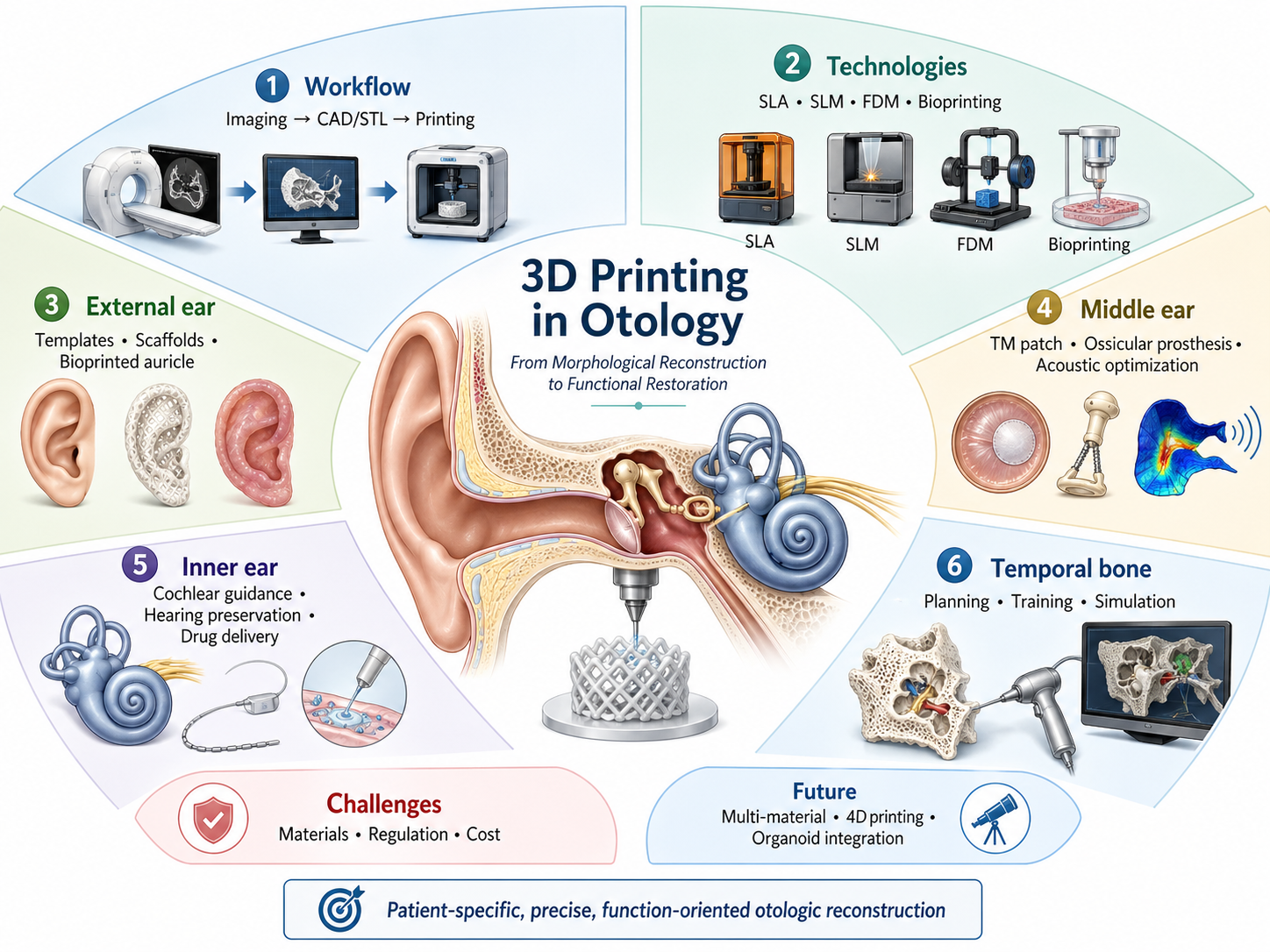
Through patient-specific design and precise anatomical fabrication, 3D printing is reshaping otology from experience-based surgery toward digital precision medicine. This review summarizes recent advances in the application of 3D printing in otology, focusing on technical platforms, morphological reconstruction, and functional restoration. At the technical level, stereolithography, selective laser melting, fused deposition modeling, and 3D bioprinting are suited to preoperative simulation, titanium-alloy implant fabrication, educational models, and tissue-engineered scaffolds, respectively. For morphological reconstruction, 3D printing enables precise replication of structures ranging from auricular frameworks and middle-ear ossicular chains to temporal-bone anatomical models. For functional restoration, patient-specific ossicular prostheses can optimize sound conduction, surgical guides can assist cochlear implantation and help preserve residual hearing, sustained-release devices may help overcome barriers to inner-ear drug delivery, and tissue-engineered scaffolds have opened new possibilities for inner-ear neural repair and regeneration. Although challenges related to material compatibility, regulatory approval, and cost-effectiveness remain, advances in multi-material printing and 4D printing may further expand the clinical potential of 3D printing in personalized otologic reconstruction that integrates anatomical form with functional recovery.
Keywords: 3D printing; otology; morphological reconstruction; functional restoration; personalized medicine
Introduction
Because of the anatomical complexity of the ear, the precision required for auditory and vestibular function, and substantial interindividual anatomical variation, otology remains one of the most technically demanding subspecialties within otolaryngology and head and neck surgery. Conditions ranging from congenital microtia and ossicular chain disruption caused by chronic otitis media to otosclerosis and sensorineural hearing loss requiring cochlear implantation all impose stringent requirements on therapeutic strategies [1]. Although conventional treatments are widely used in clinical practice, they still face substantial limitations in anatomical matching, functional restoration, operative precision, and long-term stability. Therefore, achieving accurate morphological reconstruction while maximizing auditory functional recovery has become a central challenge in contemporary otology [2].
The emergence of 3D printing offers a promising solution to this challenge. By enabling patient-specific design, precise replication of complex structures, and rapid prototyping, this technology is well suited to the highly variable anatomy of the ear. Since Kaneko [3] first applied three-dimensional shape measurement to microtia reconstruction in 1993, 3D printing in otology has evolved from the production of anatomical models to the design and fabrication of personalized implants, and further toward tissue engineering and regenerative medicine. Its applications now extend from external auricular reconstruction and middle-ear ossicular chain repair to the protection and restoration of inner-ear neural function, as well as temporal bone models for surgical training [4–5]. In this context, 3D printing is gradually reshaping otologic diagnosis and treatment by linking anatomical reconstruction with functional recovery.
This review summarizes recent progress in the application of 3D printing technology in precision otology. First, it outlines the technical foundations of 3D printing and the main application strategies in otologic practice. It then discusses the current research status and clinical value of 3D printing in outer-ear reconstruction, middle-ear ossicular chain repair, inner-ear functional restoration, and temporal bone surgical training. Finally, it summarizes the major challenges and future directions of this field, including material biocompatibility, regulatory pathways, cost-effectiveness, multi-material printing, 4D printing, and tissue-engineered reconstruction. By integrating current evidence, this review aims to provide clinicians and researchers with a structured understanding of 3D printing in otology and to support its further translational development.
Technical Workflow and Application Strategies of 3D Printing in Otology
The clinical application of 3D printing in otology relies on a complete digital-to-physical workflow that transforms patient-specific imaging data into anatomical models [6], surgical guides, implants, or tissue-engineered constructs. Because otologic structures are small, complex, and highly variable among individuals, successful application requires not only accurate image reconstruction but also careful matching between printing technology, material properties, clinical purpose, and cost-effectiveness [1]. This section therefore summarizes the technical workflow of 3D printing in otology and further discusses demand-oriented application strategies for preoperative planning, surgical guidance, personalized implantation, and regenerative reconstruction.
Digital workflow: from medical imaging to printable models
The clinical application of 3D printing begins with the conversion of patient-specific anatomical information into a printable digital model. In medical practice, anatomical data are commonly obtained from computed tomography (CT), magnetic resonance imaging (MRI), or, in selected scenarios, ultrasound imaging. These imaging datasets are usually stored in the Digital Imaging and Communications in Medicine (DICOM) format [7], which provides the basis for subsequent three-dimensional reconstruction.
After image acquisition, the target anatomical structures are segmented and reconstructed using computer-aided design (CAD) or medical image processing software. The reconstructed model is then exported into a printer-compatible file format, most commonly the Standard Tessellation Language (STL) format [8]. STL files describe the surface geometry of a three-dimensional object using triangular meshes, allowing the digital model to be sliced into multiple layers for additive manufacturing [9]. Through this workflow, patient-specific anatomical structures can be transformed from two-dimensional imaging data into physical models, surgical guides, implants, or tissue-engineered scaffolds.
In otology, this digital workflow is particularly valuable because the ear contains small, complex, and highly individualized anatomical structures. High-resolution imaging and accurate segmentation allow the reconstruction of delicate structures such as the ossicular chain, facial nerve canal, cochlea, external auricle, and temporal bone [10]. These models can support preoperative planning, surgical simulation, anatomical teaching, doctor-patient communication, and the design of personalized therapeutic devices.
Demand-oriented selection of 3D printing technologies in otology
The key to applying 3D printing in otology is not the use of a single printing method, but the selection of an appropriate technology according to clinical purpose. Different otologic applications require different levels of precision, material performance, sterilizability, fabrication time, and cost. Here are several common 3D printing methods: Stereolithography (SLA) and digital light processing (DLP) offer high precision and smooth surface finish, making them suitable for preoperative models and surgical guides, although their material selection remains limited. Selective laser melting (SLM) enables the fabrication of high-strength metal implants such as custom ossicular prostheses, but is associated with high cost and the need for post-processing. Fused deposition modeling (FDM) is low-cost and easy to operate, yet its lower precision and weak interlayer bonding restrict its use to educational models and biodegradable scaffolds. Three-dimensional bioprinting incorporates cells and growth factors for tissue-engineered auricular constructs and inner ear repair, but currently lacks mechanical strength and remains at an experimental stage (Table 1).
Table 1. Comparison of common 3D printing technologies in terms of materials, resolution, advantages, limitations, and representative otological applications.
| Technology | Typical Materials | Resolution | Advantages | Limitations | Representative Otological Applications |
|---|---|---|---|---|---|
| SLA | Photopolymer resins | ~25–100 μm | High precision, smooth surface | Limited material range, post-curing required | Preoperative models, surgical guides |
| SLM | Titanium alloys, Co–Cr alloys | ~50–200 μm | High mechanical strength, suitable for permanent implantation | High cost, surface post-processing required | Custom ossicular prostheses, auricular frameworks |
| FDM | PLA, PCL, and other thermoplastics | ~100–300 μm | Low cost, simple operation | Lower precision, weaker interlayer bonding | Educational models, biodegradable scaffolds |
| DLP | Photopolymer resins | ~50–100 μm | Fast printing speed, high precision | Limited material selection | High-resolution anatomical models |
| 3D Bioprinting | Hydrogels, bioinks | ~20–200 μm | Can incorporate cells and growth factors | Low mechanical strength, still at experimental stage | Tissue-engineered auricles, inner ear repair |
Therefore, strategies for applying 3D printing in otology can be categorized based on their roles in morphological reconstruction and functional restoration: Preoperative and educational models, as well as customized surgical guides, play a crucial role in guiding morphological reconstruction. Functional restoration includes tympanic membrane repair and targeted drug delivery, while customized implants and tissue engineering scaffolds not only enable precise morphological reconstruction but also better assist patients in restoring ear-related functions [11].
Preoperative planning and educational models
Preoperative planning models and educational models represent the earliest and most mature applications of 3D printing in otology. Their main purpose is to reproduce complex anatomical structures and spatial relationships, helping surgeons understand individual anatomical variations, anticipate surgical risks, and rehearse difficult procedures before surgery. Typical applications include temporal bone models for lateral skull base surgery [12], facial nerve decompression, congenital auricular deformity assessment [13], and resident training in otologic anatomy [14].
For these models, morphological accuracy and cost-effectiveness are more important than implantability or long-term biocompatibility. Stereolithography (SLA) and fused deposition modeling (FDM) are the most commonly used techniques. SLA provides higher printing resolution and can reproduce fine structures such as the ossicles and facial nerve canal. Resin-based SLA models also offer relatively realistic tactile feedback during drilling and are therefore useful for surgical rehearsal and detailed anatomical teaching [15]. In contrast, FDM is less expensive and more suitable for large-scale production of teaching models, basic anatomical education, and use in primary-care or resource-limited settings [16].
Patient-specific surgical guides
Surgical guides are designed to translate preoperative planning into intraoperative precision. Their core value lies in converting virtual information, including the planned entry point, direction, angle, and depth, into a physical guide that can assist surgeons during key procedural steps. In otology, such guides are mainly used in cochlear implantation, ossicular chain reconstruction, and complex temporal bone procedures [17].
Compared with ordinary anatomical models, surgical guides require higher dimensional accuracy, reliable sterilizability, close conformity to bone surfaces, and convenient intraoperative handling. SLA is currently one of the preferred techniques for fabricating surgical guides because of its high resolution and ability to generate smooth, patient-specific structures. By improving procedural accuracy and reducing intraoperative uncertainty, surgical guides may contribute to safer and more standardized otologic surgery.
Tympanic Membrane Repair and Targeted Drug Delivery to the Inner Ear
Tympanic membrane perforation is a common cause of conductive hearing loss; traditional surgical repair methods suffer from issues such as tissue harvesting trauma and poor structural compatibility. 3D printing technology offers a high-precision, personalized solution for tympanic membrane regeneration [18]. Using high-resolution 3D printing processes, scaffolds with a multi-layered biomimetic structure resembling that of the natural tympanic membrane can be fabricated [19]. In terms of material selection, current research tends to favor photo-crosslinkable natural polymers and composite hydrogels, which possess excellent biocompatibility and promote cell migration, proliferation, and tissue remodeling.
Pharmacological treatment of inner ear diseases has long been limited by the blood-labyrinth barrier and the deep anatomical location of the cochlea [20]. 3D printing technology offers a new approach to addressing this challenge, centered on the fabrication of micro-drug delivery implants that precisely conform to the round window membrane or the round window niche. Utilizing patient-specific image reconstruction, these implants can be custom-shaped to match anatomical structures, ensuring that the drug delivery device remains stably positioned at the target site. Furthermore, localized micro-delivery significantly improves drug bioavailability in the inner ear while avoiding systemic side effects, providing a novel technological platform for the precise treatment of inner ear disorders such as sensorineural hearing loss.
Customized implants for structural and functional reconstruction
Customized implants represent one of the most clinically valuable but technically demanding applications of 3D printing in otology. Their purpose is to overcome the mismatch between standardized implants and individual anatomical variations, thereby achieving personalized structural reconstruction and functional restoration. Major applications include ossicular chain reconstruction and auricular reconstruction.
For permanent implants, the key requirements include biocompatibility, mechanical stability, anatomical fit, sterilizability, and long-term safety. Selective laser melting (SLM), especially for titanium alloy fabrication, is currently one of the most important approaches for producing customized metallic implants. Titanium alloys have favorable biocompatibility and mechanical strength, making them suitable for long-term implantation [21]. In addition, 3D printing allows the fabrication of porous, hollow, or thin-walled structures, which may help reduce implant rigidity, improve tissue integration, and better mimic the mechanical properties of native ear structures.
Tissue-engineered scaffolds and 3D bioprinting
Tissue-engineered scaffolds represent the frontier of 3D printing in otology. Unlike anatomical models or inert implants, these scaffolds aim to support biological regeneration through the integrated design of biomaterials, cells, and bioactive factors [22]. Potential applications include auricular cartilage engineering, inner-ear neural repair, and local drug delivery systems.
An ideal tissue-engineered scaffold should provide cell compatibility, controlled degradation, mechanical support, appropriate porosity, and the ability to release bioactive molecules in a controlled manner [19]. 3D bioprinting offers unique advantages in this context because it can spatially organize cells and biomaterials with high precision [22]. Compared with traditional cell seeding methods, bioprinting may improve cell distribution and reduce the risk of insufficient nutrient diffusion within the scaffold. Although most applications remain at the experimental or preclinical stage, tissue-engineered scaffolds and 3D bioprinting provide an important direction for future functional reconstruction in otology.For instance, 3D-printed nanoPGA/PCL composite scaffolds have been utilized to demonstrate that optimizing scaffold surface properties through ethanol pretreatment can significantly enhance human auricular chondrocyte adhesion and promote the in vivo regeneration of mature, glossy cartilage [23] (Figure 1).
Figure 1. Design of 3D nanoPGA/PCL scaffolds seeded with human auricular chondrocytes ± ethanol treatment.
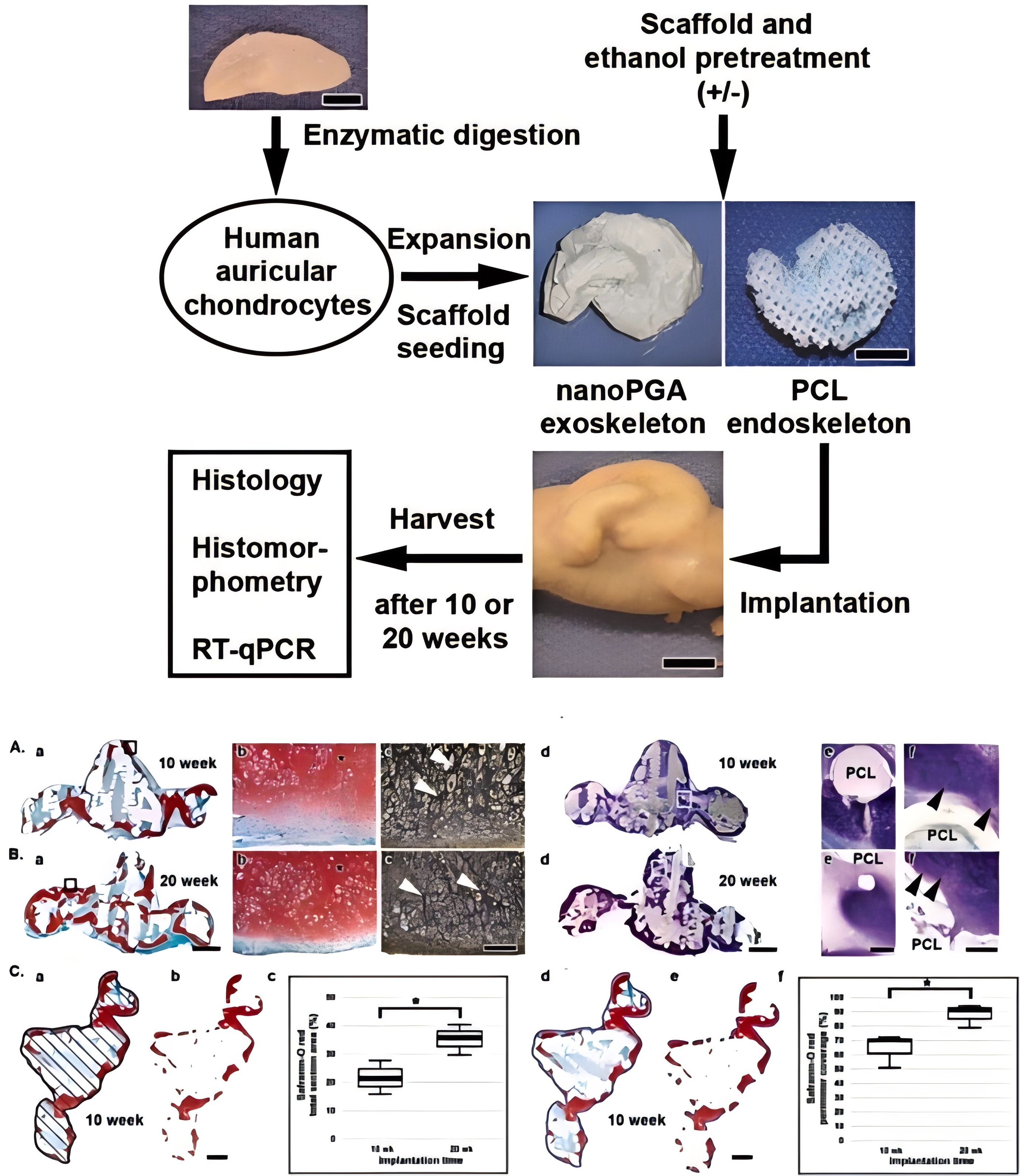
Histological analysis of regenerated cartilage on ethanol-treated scaffolds after 10 and 20 weeks of implantation [23]. Copyright 2021 PLoS One.
Clinical Applications of 3D Printing in Otology: From Morphological Reconstruction to Functional Restoration
The clinical value of 3D printing in otology lies in its ability to translate patient-specific anatomical information into tangible tools for reconstruction, simulation, implantation, and regenerative repair. Because the external ear, middle ear, inner ear, and temporal bone differ substantially in anatomical scale, functional requirements, and clinical accessibility, 3D printing strategies must be tailored to distinct therapeutic goals. In the external ear, the primary objective is to restore auricular shape and psychosocial function. In the middle ear, the key challenge is to reconstruct the ossicular chain while preserving efficient sound conduction. In the inner ear, 3D printing is increasingly used to assist cochlear implantation, improve local drug delivery, and support neural repair strategies [24]. In temporal bone surgery, printed models provide a practical platform for preoperative rehearsal and surgical education. Together, these applications indicate that 3D printing in otology is evolving from morphological replication toward function-oriented precision medicine [14].
External Ear Reconstruction: From Mirror-Image Templates to Tissue-Engineered Auricles
The external ear is important for both sound collection and facial appearance. Auricular defects, most commonly caused by congenital microtia [25], trauma, burns, or tumor resection, can impair hearing-related function and psychosocial well-being. Because the auricle has a delicate three-dimensional contour and an elastic cartilage framework, auricular reconstruction remains technically demanding [26].
Autologous rib cartilage grafting remains a standard method for auricular reconstruction because of its good biocompatibility and low immunogenicity [27]. However, it requires cartilage harvesting and precise manual sculpting, which may lead to donor-site morbidity, cartilage exposure, infection, hematoma, and variable aesthetic outcomes [20,26]. Porous polyethylene implants, such as Medpor, provide an alternative alloplastic option, but they are associated with risks such as infection, fracture, implant exposure, and limited adaptability in growing pediatric patients [27–28]. These limitations have driven the development of more individualized and less invasive reconstruction strategies.
3D printing has improved auricular reconstruction mainly by providing patient-specific templates and customized scaffolds. In unilateral microtia, imaging data from the healthy ear can be mirrored to generate a three-dimensional model of the affected side, offering surgeons a precise reference for framework sculpting, auricular positioning, and bilateral symmetry [29–30]. Compared with traditional two-dimensional templates, 3D-printed models can reduce intraoperative adjustment and improve anatomical accuracy [33]. More recently, directly printed auricular scaffolds and prostheses have been explored to reduce dependence on manual carving and improve structural consistency. However, their clinical use still requires careful evaluation of biocompatibility, mechanical elasticity, infection risk, skin coverage, and long-term stability.
Beyond inert scaffolds, 3D bioprinting and tissue engineering aim to generate living cartilage-like auricular constructs. In this strategy, chondrocytes or chondrogenic cells are combined with printable biomaterials to form bioinks, which are then printed into auricular-shaped structures and cultured to promote cartilage matrix deposition. Ideal bioinks should provide biocompatibility, tunable degradation, cell adhesion, nutrient diffusion, and cartilage-like mechanical strength [34]. Hydrogels, including collagen-, gelatin-, alginate-, and hyaluronic-acid-based systems, are attractive because of their extracellular matrix-like properties and high permeability to nutrients and soluble factors [35]. Although promising, tissue-engineered auricles remain largely experimental, and further work is needed to ensure durable shape retention, cell viability, vascularized soft-tissue coverage, and regulatory feasibility. To address these mechanical and functional limitations, recent breakthroughs have utilized stress-relaxing HATG-Alg bioinks combined with growth factor-controlled chondrogenesis to engineer functional elastic cartilage. These 3D-bioprinted constructs achieve near-native mechanical properties, such as a compressive modulus of 1.1 ± 0.03 MPa, and maintain structural integrity during in vivo evaluation [36] (Figure 2).
Figure 2. Engineering and evaluation of elastic cartilage-mimetic auricular grafts.
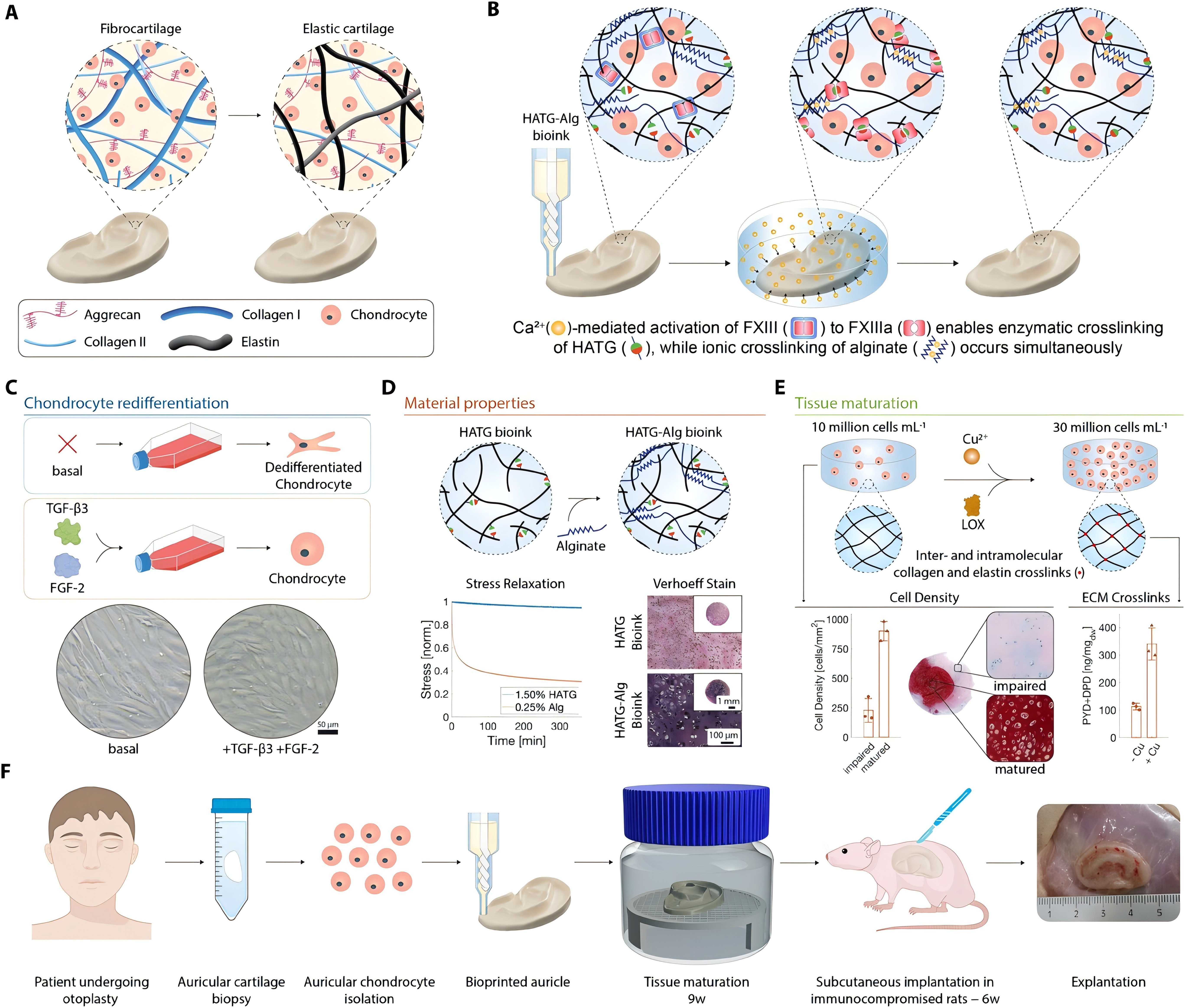
(A) Comparison of fibro- and elastic cartilage. (B) Bioink design featuring HATG and alginate crosslinking. (C-E) Strategies for promoting chondrogenic redifferentiation and tissue maturation via growth factors and LOX/Cu-mediated crosslinking. (F) Comprehensive workflow from primary cell isolation and bioprinting to in vivo subcutaneous rat model evaluation [36]. Copyright 2024 P. Fisch et al.
Middle Ear Reconstruction: Personalized Ossicular Prostheses and Acoustic Optimization
The middle ear transmits airborne sound to the cochlear fluid through the tympanic membrane and ossicular chain. Damage to the malleus, incus, or stapes caused by chronic otitis media, cholesteatoma, trauma, congenital anomalies, or surgery can disrupt sound conduction and result in conductive hearing loss. Ossicular chain reconstruction is therefore a key surgical strategy for restoring middle-ear function. However, conventional ossicular prostheses are usually produced in limited standard sizes and often require intraoperative trimming or adjustment. This may cause suboptimal prosthesis length, angular mismatch, poor fixation, displacement, extrusion, or unsatisfactory hearing outcomes [37]. Because the middle ear is a small and highly sensitive mechanical system, even minor deviations in prosthesis geometry or contact angle may affect acoustic transmission.
3D printing provides a strategy for patient-specific ossicular reconstruction. High-resolution computed tomography can be used to reconstruct the tympanic membrane, ossicular remnants, middle-ear cavity, ligaments, and surrounding structures [38]. Based on these data, customized ossicular prostheses can be designed to better fit the available anatomical space and intended reconstruction pathway. More importantly, finite element analysis can be integrated into this workflow to simulate displacement, stress, strain, and frequency response under sound stimulation [39]. This allows different prosthesis materials, shapes, lengths, and contact configurations to be evaluated before fabrication. In this sense, 3D printing is not only a manufacturing method but also part of an engineering-assisted platform for anatomical matching and acoustic optimization.
The design, 3D printing, coating, and application of GelMA patches are also currently trending technologies. Material selection remains central to the success of 3D-printed ossicular prostheses. Titanium alloys are widely used in ossiculoplasty because of their favorable biocompatibility, mechanical strength, low weight, and acoustic performance. Recent evidence suggests that titanium total ossicular replacement prostheses may achieve better postoperative air-bone gap improvement than autologous materials in total ossicular chain reconstruction [40]. 3D printing may further improve titanium-based prostheses by enabling individualized geometry, lightweight structures, and more stable coupling with residual middle-ear components. Nevertheless, 3D-printed ossicular prostheses have not yet become routine in clinical practice. Key barriers include high-resolution segmentation, manufacturing standardization, sterilization validation, cost control, and long-term clinical outcome evaluation. Future studies should integrate anatomical fit, acoustic simulation, and postoperative hearing outcomes to clarify which patient groups may benefit most from personalized 3D-printed ossicular reconstruction. Beyond ossicular chain repair, 3D printing is significantly advancing the treatment of tympanic membrane (TM) perforations. For instance, biofunctional GelMA-based patches featuring microneedles for enhanced adhesion have been fabricated using Digital Light Processing (DLP) technology. These patches can be further functionalized with antimicrobial nanoparticles via electrohydrodynamic atomization (EHDA) to prevent infection while promoting tissue regeneration, offering a highly personalized and effective approach for TM repair [41] (Figure 3).
Figure 3. Fabrication and application of 3D-printed GelMA patches for tympanic membrane repair.
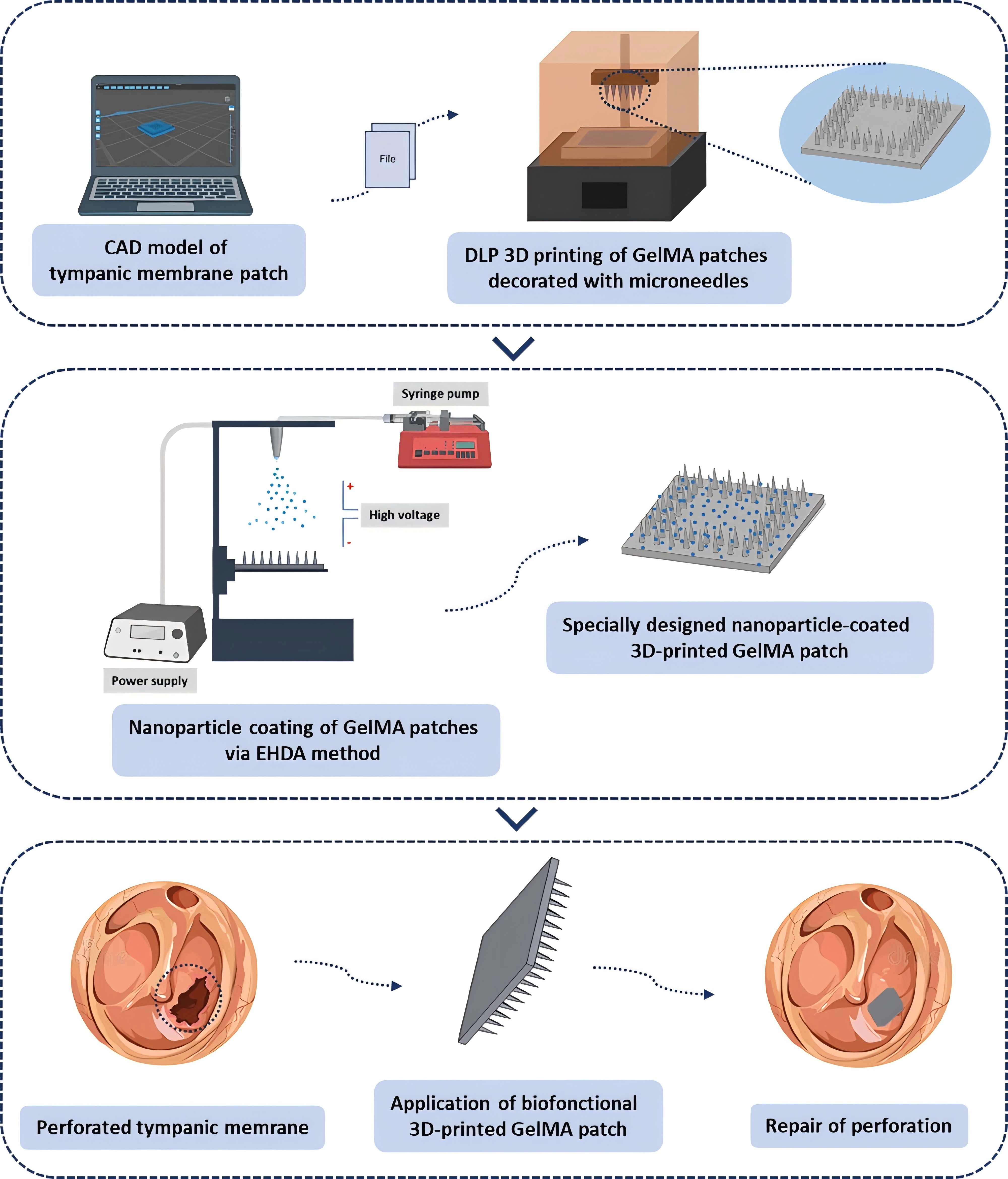
Microneedle-functionalized GelMA patches fabricated by DLP and functionalized via EHDA for antimicrobial, regenerative tympanic membrane repair [41]. Copyright 2025 T. Bedir, D. Uzun.
Inner Ear Applications: Cochlear Implantation Assistance and Local Drug Delivery
The inner ear is anatomically confined, functionally delicate, and difficult to repair once damaged [20]. Sensorineural hearing loss remains challenging to treat because hair cells and spiral ganglion neurons have limited regenerative capacity. Hearing aids are useful for mild to moderate hearing loss, whereas cochlear implants remain the most effective intervention for severe to profound sensorineural hearing loss by electrically stimulating residual spiral ganglion neurons [42]. However, cochlear implantation requires highly precise electrode placement, and preservation of residual hearing has become increasingly important as implantation indications expand. Individual variations in cochlear size, curvature, duct length, and internal anatomy may influence electrode selection, insertion depth, scalar position, and the risk of intracochlear trauma.
3D printing can support cochlear implantation by converting patient-specific imaging data into physical cochlear models for preoperative planning, electrode insertion training, and device evaluation [18]. Compared with conventional two-dimensional imaging, 3D-printed models provide a more intuitive understanding of cochlear geometry and electrode trajectory. Recent smart cochlear models with real-time feedback can further simulate electrode insertion and detect technical errors, such as excessive force, retraction, or bending [43]. One study reported that training with a smart 3D-printed cochlear model improved novice surgeons’ electrode insertion depth by 11.8% and reduced retraction and bending errors by 60.3% and 54.9%, respectively [43]. These findings suggest that 3D printing may contribute not only to anatomical visualization but also to procedural standardization and skills assessment in cochlear implantation.
Another promising application is inner-ear drug delivery. Systemic administration is limited by the blood-labyrinth barrier, whereas intratympanic injection still depends on passive diffusion across the round window membrane, making local drug concentration and exposure duration difficult to control [44]. 3D-printed microneedle-based delivery systems may provide a more precise and minimally invasive solution. Using high-resolution methods such as two-photon polymerization, micron-scale trans-tympanic or round-window devices can be fabricated to create controlled pathways for drug entry. Veit et al. developed several 3D-printed trans-tympanic delivery devices with adjustable release durations ranging from days to months [45]. These platforms may improve the delivery of ototherapeutics, including macromolecular drugs, gene therapy vectors, and neurotrophic factors. Nevertheless, inner-ear applications require strict safety evaluation because any device interacting with the round window membrane or cochlea must minimize trauma, inflammation, fibrosis, infection, and long-term hearing damage [46]. Future studies should therefore focus on biocompatibility, reversibility, delivery accuracy, and functional auditory outcomes.
Temporal Bone Models: Surgical Planning and Simulation-Based Education
Temporal bone surgery requires precise three-dimensional anatomical understanding and refined operative skills within a narrow surgical field [47]. The facial nerve, ossicles, labyrinth, sigmoid sinus, jugular bulb, carotid canal, and dura, are densely packed within a limited bony space. Traditional training has long relied on cadaveric temporal bone dissection, which remains valuable but is constrained by specimen scarcity, ethical considerations, high cost, storage requirements, non-reusability, and potential biosafety risks [49–50]. These limitations have created a strong demand for alternative platforms for preoperative planning and surgical education.
3D-printed temporal bone models have emerged as practical tools to address this need. By converting high-resolution CT data into physical models, 3D printing can reproduce patient-specific temporal bone anatomy, including complex pathological variations. In clinical practice, such models help surgeons visualize spatial relationships beyond conventional two-dimensional imaging and rehearse key steps before surgery [12], particularly in complex cases such as lateral skull base lesions, facial nerve decompression, congenital malformations, or revision procedures. In education, 3D-printed temporal bones provide a safe, reproducible, and cost-effective supplement to cadaveric specimens. They can be produced in large numbers, tailored to different levels of difficulty, and repeatedly used for structured training. Several studies have shown that these models provide acceptable anatomical fidelity and realistic depth perception, while the SAPIENS model has further demonstrated value in otologic simulation and resident training [15,51].
The utility of these models is further enhanced by advances in imaging, such as ultra-high-resolution CT and photon-counting computed tomography, which provide more detailed datasets for model reconstruction [52–53]. Nevertheless, 3D-printed temporal bone models cannot fully replace cadaveric dissection. Current limitations include incomplete simulation of soft tissue, variable drilling feedback, limited bleeding simulation, and differences in tactile realism compared with human bone. Therefore, their most appropriate role is as part of a complementary training system: 3D-printed models for early anatomical learning, repeated procedural rehearsal, and patient-specific preoperative planning, and cadaveric bones for advanced tissue handling and higher-fidelity surgical experience [53]. Such an integrated strategy may improve the accessibility, standardization, and effectiveness of otologic surgical training.
Conclusion
3D printing technology is profoundly transforming traditional models of otological diagnosis and treatment, and its unique value in precise restoration and functional reconstruction has been widely recognized. Based on the systematic review presented in this paper, research progress and future directions in this field can be summarized as follows:
At the technological foundation level, advances in imaging technology have provided high-precision data sources for 3D printing, and the application of HRCT has enabled the clear visualization of micrometer-scale ossicular chain structures; Various printing technologies each have their specific applications: SLA excels at high-precision preoperative models; SLM is the preferred process for titanium alloy implants; FDM, with its cost advantage, serves educational purposes and the preparation of biodegradable scaffolds; and 3D bioprinting offers entirely new possibilities for tissue-engineered auricles; material selection strategies are becoming increasingly sophisticated.
In terms of morphological reconstruction, 3D printing technology has achieved comprehensive coverage from the outer ear to the middle ear. In the field of outer ear reconstruction, advancements range from Kelly’s 3D template model in 1993 to today’s personalized titanium alloy scaffolds; in middle ear reconstruction, personalized ossicular prostheses have resolved the core issue of size mismatch in traditional prostheses through precise anatomical alignment; and temporal bone anatomical models have opened new avenues for resident training and preoperative simulation of complex cases, effectively addressing the shortage of cadaveric specimens.
However, the clinical translation of 3D printing technology in otology still faces multiple challenges. In terms of materials science, existing materials struggle to simultaneously meet the dual requirements of mechanical support and biocompatibility; regarding regulatory frameworks, the approval pathway for personalized implants remains unclear, and quality control standards have yet to be established; in terms of clinical adoption, printing costs and time efficiency remain significant barriers to widespread application. Resolving these issues requires the deep integration of materials science, biomedical engineering, clinical medicine, and regulatory science.
Looking ahead, with the development of cutting-edge technologies such as 4D printing, multi-material printing, and organ-on-a-chip, 3D printing in otology will continue to evolve toward intelligence, functionality, and personalization. The application of shape-memory materials holds promise for the development of smart implants that automatically unfold after minimally invasive implantation; multi-material gradient printing can construct functional scaffolds with gradually varying mechanical properties and bioactivity; and the combination of organoid technology with 3D printing may offer entirely new therapeutic strategies for intractable diseases such as inner ear hair cell regeneration. From the first 3D-printed ear template in 1993 to today’s bioprinted auricles [3], over three decades of development have witnessed this technology’s transformation from a “supporting tool” to a “therapeutic modality.” We have every reason to believe that, with the deepening advancement of interdisciplinary collaboration, 3D printing technology will inevitably propel otological diagnosis and treatment into a new era of personalized precision medicine that unifies form and function.
Abbreviations
3D: three-dimensional; 4D: four-dimensional; CAD: computer-aided design; CT: computed tomography; DICOM: Digital Imaging and Communications in Medicine; DLP: Digital Light Processing; ECM: Extracellular matrix; EHDA: Electrohydrodynamic Atomization; FDM: fused deposition modeling; FGF-2: Fibroblast growth factor 2; FXIII: Factor XIII; GelMA: Gelatin Methacryloyl; HATG: Heparin-conjugated thrombin-activatable transglutaminase; HRCT: High-Resolution Computed Tomography; LOX: Lysyl oxidase; MRI: Magnetic Resonance Imaging; PCL: Polycaprolactone; PLA: Polylactic Acid; SLA: Stereolithography; SLM: Selective Laser Melting; STL: Standard Tessellation Language; TGF-β3: Transforming Growth Factor Beta 3.
Declarations
Author Contributions
HYB and ZHY supervised the study and critically revised the manuscript. All authors read and approved the final manuscript.
Ethical Approval
Not applicable.
Funding
This research received no external funding.
Competing Interests
The authors declare no competing interests.
Data Availability
All data generated or analyzed during this study are included in this published article.
Acknowledgments
We would like to thank the anonymous reviewers and the editors for their constructive comments.
References
[1] M. Stavrakas et al. , ‘3D Printing in ENT Training: Current State-of-the-Art and Perspectives’, Maedica , vol. 19, no. 3, Sep. 2024, https://doi.org/10.26574/maedica.2024.19.3.629
[2] F. Zoccali et al. , ‘3D Printing in Otolaryngology Surgery: Descriptive Review of Literature to Define the State of the Art’, Healthcare , vol. 11, no. 1, p. 108, Dec. 2022, https://doi.org/10.3390/healthcare11010108
[3] T. Kaneko, ‘A System for Three-Dimensional Shape Measurement and its Application in Microtia Ear Reconstruction.’, Keio j. med , vol. 42, no. 1, pp. 22–40, 1993, https://doi.org/10.2302/kjm.42.22
[4] N. Zhong and X. Zhao, ‘3D printing for clinical application in otorhinolaryngology’, Eur Arch Otorhinolaryngol , vol. 274, no. 12, pp. 4079–4089, Dec. 2017, https://doi.org/10.1007/s00405-017-4743-0
[5] M. A. De Souza, R. F. Bento, P. T. Lopes, D. M. De Pinto Rangel, and L. Formighieri, ‘Three-dimensional printing in otolaryngology education: a systematic review’, Eur Arch Otorhinolaryngol , vol. 279, no. 4, pp. 1709–1719, Apr. 2022, https://doi.org/10.1007/s00405-021-07088-7
[6] L. F. Velásquez-García and Y. Kornbluth, ‘Biomedical Applications of Metal 3D Printing’, Annu. Rev. Biomed. Eng. , vol. 23, no. 1, pp. 307–338, Jul. 2021, https://doi.org/10.1146/annurev-bioeng-082020-032402
[7] M. Larobina, ‘Thirty Years of the DICOM Standard’, Tomography , vol. 9, no. 5, pp. 1829–1838, Oct. 2023, https://doi.org/10.3390/tomography9050145
[8] B. C. Gross, J. L. Erkal, S. Y. Lockwood, C. Chen, and D. M. Spence, ‘Evaluation of 3D Printing and Its Potential Impact on Biotechnology and the Chemical Sciences’, Anal. Chem. , vol. 86, no. 7, pp. 3240–3253, Apr. 2014, https://doi.org/10.1021/ac403397r
[9] M. Pérez-Sevilla, F. Rivas-Navazo, P. Latorre-Carmona, and D. Fernández-Zoppino, ‘Protocol for Converting DICOM Files to STL Models Using 3D Slicer and Ultimaker Cura’, JPM , vol. 15, no. 3, p. 118, Mar. 2025, https://doi.org/10.3390/jpm15030118
[10] A. Marro, T. Bandukwala, and W. Mak, ‘Three-Dimensional Printing and Medical Imaging: A Review of the Methods and Applications’, Current Problems in Diagnostic Radiology , vol. 45, no. 1, pp. 2–9, Jan. 2016, https://doi.org/10.1067/j.cpradiol.2015.07.009
[11] A. Omari, M. Frendø, M. S. Sørensen, S. A. W. Andersen, and A. Frithioff, ‘The cutting edge of customized surgery: 3D-printed models for patient-specific interventions in otology and auricular management—a systematic review’, Eur Arch Otorhinolaryngol , vol. 279, no. 7, pp. 3269–3288, Jul. 2022, https://doi.org/10.1007/s00405-022-07291-0
[12] A. G. Micco, ‘Are Simulated or 3D Printed Temporal Bones as Useful for Training as Cadaveric Bones’, Laryngoscope , vol. 133, no. 8, pp. 1782–1783, Aug. 2023, https://doi.org/10.1002/lary.30704
[13] C. L. Reighard, S. J. Hollister, and D. A. Zopf, ‘Auricular reconstruction from rib to 3D printing’, J 3D Print Med , vol. 2, no. 1, pp. 35–41, Jan. 2018, https://doi.org/10.2217/3dp-2017-0017
[14] A. Frithioff, M. Frendø, D. B. Pedersen, M. S. Sørensen, and S. A. Wuyts Andersen, ‘3D‐Printed Models for Temporal Bone Surgical Training: A Systematic Review’, Otolaryngol.--head neck surg. , vol. 165, no. 5, pp. 617–625, Nov. 2021, https://doi.org/10.1177/0194599821993384
[15] G. Iannella et al. , ‘The SAPIENS 3D-printed temporal bone model: a real tool for advanced otologic surgery education’, Eur Arch Otorhinolaryngol , vol. 282, no. 6, pp. 3051–3063, Jun. 2025, https://doi.org/10.1007/s00405-024-09199-3
[16] K. Takahashi et al. , ‘Creating an Optimal 3D Printed Model for Temporal Bone Dissection Training’, Ann Otol Rhinol Laryngol , vol. 126, no. 7, pp. 530–536, Jul. 2017, https://doi.org/10.1177/0003489417705395
[17] L. M. Markodimitraki, T. C. Ten Harkel, R. L. A. W. Bleys, I. Stegeman, and H. G. X. M. Thomeer, ‘Cochlear implant positioning and fixation using 3D-printed patient specific surgical guides; a cadaveric study’, PLoS ONE , vol. 17, no. 7, p. e0270517, Jul. 2022, https://doi.org/10.1371/journal.pone.0270517
[18] A. Torrisi et al. , ‘The Promise and Challenges of 3D Bioprinting in Otolaryngology: A Contemporary Perspective Viewpoint’, Clinical Otolaryngology , vol. 50, no. 5, pp. 795–807, Sep. 2025, https://doi.org/10.1111/coa.14333
[19] Y. Huang et al. , ‘The application and progress of tissue engineering and biomaterial scaffolds for total auricular reconstruction in microtia’, Front. Bioeng. Biotechnol. , vol. 11, p. 1089031, Sep. 2023, https://doi.org/10.3389/fbioe.2023.1089031
[20] S.-W. Wu, Z.-Z. Nian, W. Lin, and X.-D. Zhang, ‘Unveiling the Intricacies of the Inner Ear Anatomy: Novel 3D-Printed Model for Detailed Visualization and Functional Demonstrations’, J. Laryngol. Otol. , vol. 138, no. 7, pp. 787–791, Jul. 2024, https://doi.org/10.1017/S0022215124000367
[21] W. Zhang et al. , ‘Functional hydrogel empowering 3D printing titanium alloys’, Materials Today Bio , vol. 30, p. 101422, Feb. 2025, https://doi.org/10.1016/j.mtbio.2024.101422
[22] A. McMillan, N. McMillan, N. Gupta, S. P. Kanotra, and A. K. Salem, ‘3D Bioprinting in Otolaryngology: A Review’, Adv Healthcare Materials , vol. 12, no. 19, p. 2203268, Jul. 2023, https://doi.org/10.1002/adhm.202203268
[23] N. Hirano et al. , ‘Ethanol treatment of nanoPGA/PCL composite scaffolds enhances human chondrocyte development in the cellular microenvironment of tissue-engineered auricle constructs’, PLOS ONE , vol. 16, no. 7, p. e0253149, Jul. 2021, https://doi.org/10.1371/journal.pone.0253149
[24] T. D. Crafts, S. E. Ellsperman, T. J. Wannemuehler, T. D. Bellicchi, T. Z. Shipchandler, and A. V. Mantravadi, ‘Three-Dimensional Printing and Its Applications in Otorhinolaryngology-Head and Neck Surgery’, Otolaryngol Head Neck Surg , vol. 156, no. 6, pp. 999–1010, Jun. 2017, https://doi.org/10.1177/0194599816678372
[25] D. V. Luquetti, C. L. Heike, A. V. Hing, M. L. Cunningham, and T. C. Cox, ‘Microtia: epidemiology and genetics’, Am J Med Genet A , vol. 158A, no. 1, pp. 124–139, Jan. 2012, https://doi.org/10.1002/ajmg.a.34352
[26] Y. Guo et al. , ‘Innovative tissue engineering strategies for auricular regeneration in microtia: Current advances and future perspectives’, Regenerative Therapy , vol. 30, pp. 665–680, Dec. 2025, https://doi.org/10.1016/j.reth.2025.08.010
[27] R. C. Tanzer, ‘TOTAL RECONSTRUCTION OF THE EXTERNAL EAR’:, Plastic and Reconstructive Surgery , vol. 23, no. 1, pp. 1–15, Jan. 1959, https://doi.org/10.1097/00006534-195901000-00001
[28] J. A. Cabin, M. Bassiri-Tehrani, A. P. Sclafani, and T. Romo, ‘Microtia Reconstruction’, Facial Plastic Surgery Clinics of North America , vol. 22, no. 4, pp. 623–638, Nov. 2014, https://doi.org/10.1016/j.fsc.2014.07.004
[29] R. Cenzi, A. Farina, L. Zuccarino, and F. Carinci, ‘Clinical outcome of 285 Medpor grafts used for craniofacial reconstruction’, J Craniofac Surg , vol. 16, no. 4, pp. 526–530, Jul. 2005, https://doi.org/10.1097/01.scs.0000168761.46700.dc
[30] J. D. Williams, T. Romo, A. P. Sclafani, and H. Cho, ‘Porous High-Density Polyethylene Implants in Auricular Reconstruction’, Archives of Otolaryngology - Head and Neck Surgery , vol. 123, no. 6, pp. 578–583, Jun. 1997, https://doi.org/10.1001/archotol.1997.01900060020003
[31] J. Y. Park, Y.-J. Choi, J.-H. Shim, J. H. Park, and D.-W. Cho, ‘Development of a 3D cell printed structure as an alternative to autologs cartilage for auricular reconstruction’, J Biomed Mater Res B Appl Biomater , vol. 105, no. 5, pp. 1016–1028, Jul. 2017, https://doi.org/10.1002/jbm.b.33639
[32] H.-Y. Chen, L.-S. Ng, C.-S. Chang, T.-C. Lu, N.-H. Chen, and Z.-C. Chen, ‘Pursuing Mirror Image Reconstruction in Unilateral Microtia: Customizing Auricular Framework by Application of Three-Dimensional Imaging and Three-Dimensional Printing’, Plast Reconstr Surg , vol. 139, no. 6, pp. 1433–1443, Jun. 2017, https://doi.org/10.1097/PRS.0000000000003374
[33] Y. Deng et al. , ‘Three-dimensional printing technology for the positioning of the reconstructed auricle in microtia reconstruction’, Journal of Stomatology Oral and Maxillofacial Surgery , vol. 126, no. 4, p. 102207, Sep. 2025, https://doi.org/10.1016/j.jormas.2024.102207
[34] S. Gao et al. , ‘3D printing tissue-engineered scaffolds for auricular reconstruction’, Mater Today Bio , vol. 27, p. 101141, Jul. 2024, https://doi.org/10.1016/j.mtbio.2024.101141
[35] H. Wang et al. , ‘Chondrocyte-laden gelatin/sodium alginate hydrogel integrating 3D printed PU scaffold for auricular cartilage reconstruction’, International Journal of Biological Macromolecules , vol. 253, p. 126294, Dec. 2023, https://doi.org/10.1016/j.ijbiomac.2023.126294
[36] P. Fisch et al. , ‘Tissue Engineered Human Elastic Cartilage From Primary Auricular Chondrocytes for Ear Reconstruction’, Advanced Functional Materials , vol. n/a, no. n/a, p. e30253, https://doi.org/10.1002/adfm.202530253
[37] M. Mohseni-Dargah, C. Pastras, P. Mukherjee, K. Cheng, K. Khajeh, and M. Asadnia, ‘Finite element analysis of anatomically-modelled prosthetic incus for ossicular chain reconstruction’, Comput Biol Med , vol. 187, p. 109770, Mar. 2025, https://doi.org/10.1016/j.compbiomed.2025.109770
[38] T. Fuse, M. Aoyagi, Y. Koike, and Y. Sugai, ‘Diagnosis of the Ossicular Chain in the Middle Ear by High-Resolution CT’, ORL , vol. 54, no. 5, pp. 251–254, 1992, https://doi.org/10.1159/000276308
[39] H. Mohammadi, A. Ebrahimian, and N. Maftoon, ‘Finite-element modelling of interactions of needle with tympanic membrane and middle ear’, Hearing Research , vol. 452, p. 109092, Oct. 2024, https://doi.org/10.1016/j.heares.2024.109092
[40] N. S. Andresen, F. Kostadinov, C. Schlegel, and T. Linder, ‘Total Ossicular Chain Reconstruction: Is Autologous Material Better Than a TORP?’, Otology & Neurotology , vol. 47, no. 3, pp. 511–516, Mar. 2026, https://doi.org/10.1097/MAO.0000000000004782
[41] T. Bedir et al. , ‘Development of Specially Designed Nanoparticle-coated 3D-printed Gelatin Methacryloyl Patches for Potential Tissue Engineering Applications’, Macromolecular Materials and Engineering , vol. 310, no. 2, p. 2400199, 2025, https://doi.org/10.1002/mame.202400199
[42] K. C. Schvartz-Leyzac, D. J. Colesa, D. L. Swiderski, Y. Raphael, and B. E. Pfingst, ‘Cochlear Health and Cochlear-implant Function’, JARO , vol. 24, no. 1, pp. 5–29, Jan. 2023, https://doi.org/10.1007/s10162-022-00882-y
[43] I. M. Lei et al. , ‘3D printed biomimetic cochleae and machine learning co-modelling provides clinical informatics for cochlear implant patients’, Nat Commun , vol. 12, no. 1, p. 6260, Oct. 2021, https://doi.org/10.1038/s41467-021-26491-6
[44] X. Maimaitikelimu, Z. Xuan, H. Ren, K. Chen, H. Zhang, and H. Wang, ‘Rational Design of Inner Ear Drug Delivery Systems’, Adv Sci (Weinh) , vol. 12, no. 29, p. 2410568, May 2025, https://doi.org/10.1002/advs.202410568
[45] J. G. S. Veit et al. , ‘Microneedle Designs for Trans-Tympanic Delivery of Topical Ototherapeutics’, ACS Biomater. Sci. Eng. , vol. 11, no. 12, pp. 7282–7293, Dec. 2025, https://doi.org/10.1021/acsbiomaterials.5c01689
[46] Z. Yi, X. Wang, G. Yin, and Y. Sun, ‘The Blood-Labyrinth Barrier: Non-Invasive Delivery Strategies for Inner Ear Drug Delivery’, Pharmaceutics , vol. 17, no. 4, p. 482, Apr. 2025, https://doi.org/10.3390/pharmaceutics17040482
[47] M. E. Freiser, A. Ghodadra, A. A. McCall, A. D. Shaffer, M. Magnetta, and N. Jabbour, ‘Operable, Low-Cost, High-Resolution, Patient-Specific 3D Printed Temporal Bones for Surgical Simulation and Evaluation’, Ann Otol Rhinol Laryngol , vol. 130, no. 9, pp. 1044–1051, Sep. 2021, https://doi.org/10.1177/0003489421993733
[48] B. G. Fennessy and P. O’Sullivan, ‘Establishing a temporal bone laboratory: considerations for ENT specialist training’, Ir J Med Sci , vol. 178, no. 4, pp. 393–395, Dec. 2009, https://doi.org/10.1007/s11845-009-0373-x
[49] S. M. Naik, M. S. Naik, and N. K. Bains, ‘Cadaveric temporal bone dissection: is it obsolete today?’, Int Arch Otorhinolaryngol , vol. 18, no. 1, pp. 63–67, Jan. 2014, https://doi.org/10.1055/s-0033-1351681
[50] G. Iannella et al. , ‘A new 3D-printed temporal bone: ’the SAPIENS’-specific anatomical printed-3D-model in education and new surgical simulations’, Eur Arch Otorhinolaryngol , vol. 281, no. 9, pp. 4617–4626, Sep. 2024, https://doi.org/10.1007/s00405-024-08645-6
[51] D. Pellby et al. , ‘Feasibility of micro-computed tomography for evaluating anatomical landmarks in temporal bone specimens’, Radiation Protection Dosimetry , vol. 202, no. 3–4, pp. 180–186, Mar. 2026, https://doi.org/10.1093/rpd/ncaf146
[52] F. Boubaker et al. , ‘Ultra-high-resolution CT of the temporal bone: Technical aspects, current applications and future directions’, Diagnostic and Interventional Imaging , vol. 106, no. 6, pp. 205–216, Jun. 2025, https://doi.org/10.1016/j.diii.2025.02.003
[53] W. W. Chien, M. J. da Cruz, and H. W. Francis, ‘Validation of a 3D-printed human temporal bone model for otology surgical skill training’, World J Otorhinolaryngol Head Neck Surg , vol. 7, no. 2, pp. 88–93, Jan. 2021, https://doi.org/10.1016/j.wjorl.2020.12.004
 Figures
 References
 Peer
 Information
Figure 1. Design of 3D nanoPGA/PCL scaffolds seeded with human auricular chondrocytes ± ethanol treatment.
Histological analysis of regenerated cartilage on ethanol-treated scaffolds after 10 and 20 weeks of implantation [23]. Copyright 2021 PLoS One.
Figure 2. Engineering and evaluation of elastic cartilage-mimetic auricular grafts.
(A) Comparison of fibro- and elastic cartilage. (B) Bioink design featuring HATG and alginate crosslinking. (C-E) Strategies for promoting chondrogenic redifferentiation and tissue maturation via growth factors and LOX/Cu-mediated crosslinking. (F) Comprehensive workflow from primary cell isolation and bioprinting to in vivo subcutaneous rat model evaluation [36]. Copyright 2024 P. Fisch et al.
Figure 3. Fabrication and application of 3D-printed GelMA patches for tympanic membrane repair.
Microneedle-functionalized GelMA patches fabricated by DLP and functionalized via EHDA for antimicrobial, regenerative tympanic membrane repair [41]. Copyright 2025 T. Bedir, D. Uzun.
[1] M. Stavrakas et al. , ‘3D Printing in ENT Training: Current State-of-the-Art and Perspectives’, Maedica , vol. 19, no. 3, Sep. 2024, https://doi.org/10.26574/maedica.2024.19.3.629
[2] F. Zoccali et al. , ‘3D Printing in Otolaryngology Surgery: Descriptive Review of Literature to Define the State of the Art’, Healthcare , vol. 11, no. 1, p. 108, Dec. 2022, https://doi.org/10.3390/healthcare11010108
[3] T. Kaneko, ‘A System for Three-Dimensional Shape Measurement and its Application in Microtia Ear Reconstruction.’, Keio j. med , vol. 42, no. 1, pp. 22–40, 1993, https://doi.org/10.2302/kjm.42.22
[4] N. Zhong and X. Zhao, ‘3D printing for clinical application in otorhinolaryngology’, Eur Arch Otorhinolaryngol , vol. 274, no. 12, pp. 4079–4089, Dec. 2017, https://doi.org/10.1007/s00405-017-4743-0
[5] M. A. De Souza, R. F. Bento, P. T. Lopes, D. M. De Pinto Rangel, and L. Formighieri, ‘Three-dimensional printing in otolaryngology education: a systematic review’, Eur Arch Otorhinolaryngol , vol. 279, no. 4, pp. 1709–1719, Apr. 2022, https://doi.org/10.1007/s00405-021-07088-7
[6] L. F. Velásquez-García and Y. Kornbluth, ‘Biomedical Applications of Metal 3D Printing’, Annu. Rev. Biomed. Eng. , vol. 23, no. 1, pp. 307–338, Jul. 2021, https://doi.org/10.1146/annurev-bioeng-082020-032402
[7] M. Larobina, ‘Thirty Years of the DICOM Standard’, Tomography , vol. 9, no. 5, pp. 1829–1838, Oct. 2023, https://doi.org/10.3390/tomography9050145
[8] B. C. Gross, J. L. Erkal, S. Y. Lockwood, C. Chen, and D. M. Spence, ‘Evaluation of 3D Printing and Its Potential Impact on Biotechnology and the Chemical Sciences’, Anal. Chem. , vol. 86, no. 7, pp. 3240–3253, Apr. 2014, https://doi.org/10.1021/ac403397r
[9] M. Pérez-Sevilla, F. Rivas-Navazo, P. Latorre-Carmona, and D. Fernández-Zoppino, ‘Protocol for Converting DICOM Files to STL Models Using 3D Slicer and Ultimaker Cura’, JPM , vol. 15, no. 3, p. 118, Mar. 2025, https://doi.org/10.3390/jpm15030118
[10] A. Marro, T. Bandukwala, and W. Mak, ‘Three-Dimensional Printing and Medical Imaging: A Review of the Methods and Applications’, Current Problems in Diagnostic Radiology , vol. 45, no. 1, pp. 2–9, Jan. 2016, https://doi.org/10.1067/j.cpradiol.2015.07.009
[11] A. Omari, M. Frendø, M. S. Sørensen, S. A. W. Andersen, and A. Frithioff, ‘The cutting edge of customized surgery: 3D-printed models for patient-specific interventions in otology and auricular management—a systematic review’, Eur Arch Otorhinolaryngol , vol. 279, no. 7, pp. 3269–3288, Jul. 2022, https://doi.org/10.1007/s00405-022-07291-0
[12] A. G. Micco, ‘Are Simulated or 3D Printed Temporal Bones as Useful for Training as Cadaveric Bones’, Laryngoscope , vol. 133, no. 8, pp. 1782–1783, Aug. 2023, https://doi.org/10.1002/lary.30704
[13] C. L. Reighard, S. J. Hollister, and D. A. Zopf, ‘Auricular reconstruction from rib to 3D printing’, J 3D Print Med , vol. 2, no. 1, pp. 35–41, Jan. 2018, https://doi.org/10.2217/3dp-2017-0017
[14] A. Frithioff, M. Frendø, D. B. Pedersen, M. S. Sørensen, and S. A. Wuyts Andersen, ‘3D‐Printed Models for Temporal Bone Surgical Training: A Systematic Review’, Otolaryngol.--head neck surg. , vol. 165, no. 5, pp. 617–625, Nov. 2021, https://doi.org/10.1177/0194599821993384
[15] G. Iannella et al. , ‘The SAPIENS 3D-printed temporal bone model: a real tool for advanced otologic surgery education’, Eur Arch Otorhinolaryngol , vol. 282, no. 6, pp. 3051–3063, Jun. 2025, https://doi.org/10.1007/s00405-024-09199-3
[16] K. Takahashi et al. , ‘Creating an Optimal 3D Printed Model for Temporal Bone Dissection Training’, Ann Otol Rhinol Laryngol , vol. 126, no. 7, pp. 530–536, Jul. 2017, https://doi.org/10.1177/0003489417705395
[17] L. M. Markodimitraki, T. C. Ten Harkel, R. L. A. W. Bleys, I. Stegeman, and H. G. X. M. Thomeer, ‘Cochlear implant positioning and fixation using 3D-printed patient specific surgical guides; a cadaveric study’, PLoS ONE , vol. 17, no. 7, p. e0270517, Jul. 2022, https://doi.org/10.1371/journal.pone.0270517
[18] A. Torrisi et al. , ‘The Promise and Challenges of 3D Bioprinting in Otolaryngology: A Contemporary Perspective Viewpoint’, Clinical Otolaryngology , vol. 50, no. 5, pp. 795–807, Sep. 2025, https://doi.org/10.1111/coa.14333
[19] Y. Huang et al. , ‘The application and progress of tissue engineering and biomaterial scaffolds for total auricular reconstruction in microtia’, Front. Bioeng. Biotechnol. , vol. 11, p. 1089031, Sep. 2023, https://doi.org/10.3389/fbioe.2023.1089031
[20] S.-W. Wu, Z.-Z. Nian, W. Lin, and X.-D. Zhang, ‘Unveiling the Intricacies of the Inner Ear Anatomy: Novel 3D-Printed Model for Detailed Visualization and Functional Demonstrations’, J. Laryngol. Otol. , vol. 138, no. 7, pp. 787–791, Jul. 2024, https://doi.org/10.1017/S0022215124000367
[21] W. Zhang et al. , ‘Functional hydrogel empowering 3D printing titanium alloys’, Materials Today Bio , vol. 30, p. 101422, Feb. 2025, https://doi.org/10.1016/j.mtbio.2024.101422
[22] A. McMillan, N. McMillan, N. Gupta, S. P. Kanotra, and A. K. Salem, ‘3D Bioprinting in Otolaryngology: A Review’, Adv Healthcare Materials , vol. 12, no. 19, p. 2203268, Jul. 2023, https://doi.org/10.1002/adhm.202203268
[23] N. Hirano et al. , ‘Ethanol treatment of nanoPGA/PCL composite scaffolds enhances human chondrocyte development in the cellular microenvironment of tissue-engineered auricle constructs’, PLOS ONE , vol. 16, no. 7, p. e0253149, Jul. 2021, https://doi.org/10.1371/journal.pone.0253149
[24] T. D. Crafts, S. E. Ellsperman, T. J. Wannemuehler, T. D. Bellicchi, T. Z. Shipchandler, and A. V. Mantravadi, ‘Three-Dimensional Printing and Its Applications in Otorhinolaryngology-Head and Neck Surgery’, Otolaryngol Head Neck Surg , vol. 156, no. 6, pp. 999–1010, Jun. 2017, https://doi.org/10.1177/0194599816678372
[25] D. V. Luquetti, C. L. Heike, A. V. Hing, M. L. Cunningham, and T. C. Cox, ‘Microtia: epidemiology and genetics’, Am J Med Genet A , vol. 158A, no. 1, pp. 124–139, Jan. 2012, https://doi.org/10.1002/ajmg.a.34352
[26] Y. Guo et al. , ‘Innovative tissue engineering strategies for auricular regeneration in microtia: Current advances and future perspectives’, Regenerative Therapy , vol. 30, pp. 665–680, Dec. 2025, https://doi.org/10.1016/j.reth.2025.08.010
[27] R. C. Tanzer, ‘TOTAL RECONSTRUCTION OF THE EXTERNAL EAR’:, Plastic and Reconstructive Surgery , vol. 23, no. 1, pp. 1–15, Jan. 1959, https://doi.org/10.1097/00006534-195901000-00001
[28] J. A. Cabin, M. Bassiri-Tehrani, A. P. Sclafani, and T. Romo, ‘Microtia Reconstruction’, Facial Plastic Surgery Clinics of North America , vol. 22, no. 4, pp. 623–638, Nov. 2014, https://doi.org/10.1016/j.fsc.2014.07.004
[29] R. Cenzi, A. Farina, L. Zuccarino, and F. Carinci, ‘Clinical outcome of 285 Medpor grafts used for craniofacial reconstruction’, J Craniofac Surg , vol. 16, no. 4, pp. 526–530, Jul. 2005, https://doi.org/10.1097/01.scs.0000168761.46700.dc
[30] J. D. Williams, T. Romo, A. P. Sclafani, and H. Cho, ‘Porous High-Density Polyethylene Implants in Auricular Reconstruction’, Archives of Otolaryngology - Head and Neck Surgery , vol. 123, no. 6, pp. 578–583, Jun. 1997, https://doi.org/10.1001/archotol.1997.01900060020003
[31] J. Y. Park, Y.-J. Choi, J.-H. Shim, J. H. Park, and D.-W. Cho, ‘Development of a 3D cell printed structure as an alternative to autologs cartilage for auricular reconstruction’, J Biomed Mater Res B Appl Biomater , vol. 105, no. 5, pp. 1016–1028, Jul. 2017, https://doi.org/10.1002/jbm.b.33639
[32] H.-Y. Chen, L.-S. Ng, C.-S. Chang, T.-C. Lu, N.-H. Chen, and Z.-C. Chen, ‘Pursuing Mirror Image Reconstruction in Unilateral Microtia: Customizing Auricular Framework by Application of Three-Dimensional Imaging and Three-Dimensional Printing’, Plast Reconstr Surg , vol. 139, no. 6, pp. 1433–1443, Jun. 2017, https://doi.org/10.1097/PRS.0000000000003374
[33] Y. Deng et al. , ‘Three-dimensional printing technology for the positioning of the reconstructed auricle in microtia reconstruction’, Journal of Stomatology Oral and Maxillofacial Surgery , vol. 126, no. 4, p. 102207, Sep. 2025, https://doi.org/10.1016/j.jormas.2024.102207
[34] S. Gao et al. , ‘3D printing tissue-engineered scaffolds for auricular reconstruction’, Mater Today Bio , vol. 27, p. 101141, Jul. 2024, https://doi.org/10.1016/j.mtbio.2024.101141
[35] H. Wang et al. , ‘Chondrocyte-laden gelatin/sodium alginate hydrogel integrating 3D printed PU scaffold for auricular cartilage reconstruction’, International Journal of Biological Macromolecules , vol. 253, p. 126294, Dec. 2023, https://doi.org/10.1016/j.ijbiomac.2023.126294
[36] P. Fisch et al. , ‘Tissue Engineered Human Elastic Cartilage From Primary Auricular Chondrocytes for Ear Reconstruction’, Advanced Functional Materials , vol. n/a, no. n/a, p. e30253, https://doi.org/10.1002/adfm.202530253
[37] M. Mohseni-Dargah, C. Pastras, P. Mukherjee, K. Cheng, K. Khajeh, and M. Asadnia, ‘Finite element analysis of anatomically-modelled prosthetic incus for ossicular chain reconstruction’, Comput Biol Med , vol. 187, p. 109770, Mar. 2025, https://doi.org/10.1016/j.compbiomed.2025.109770
[38] T. Fuse, M. Aoyagi, Y. Koike, and Y. Sugai, ‘Diagnosis of the Ossicular Chain in the Middle Ear by High-Resolution CT’, ORL , vol. 54, no. 5, pp. 251–254, 1992, https://doi.org/10.1159/000276308
[39] H. Mohammadi, A. Ebrahimian, and N. Maftoon, ‘Finite-element modelling of interactions of needle with tympanic membrane and middle ear’, Hearing Research , vol. 452, p. 109092, Oct. 2024, https://doi.org/10.1016/j.heares.2024.109092
[40] N. S. Andresen, F. Kostadinov, C. Schlegel, and T. Linder, ‘Total Ossicular Chain Reconstruction: Is Autologous Material Better Than a TORP?’, Otology & Neurotology , vol. 47, no. 3, pp. 511–516, Mar. 2026, https://doi.org/10.1097/MAO.0000000000004782
[41] T. Bedir et al. , ‘Development of Specially Designed Nanoparticle-coated 3D-printed Gelatin Methacryloyl Patches for Potential Tissue Engineering Applications’, Macromolecular Materials and Engineering , vol. 310, no. 2, p. 2400199, 2025, https://doi.org/10.1002/mame.202400199
[42] K. C. Schvartz-Leyzac, D. J. Colesa, D. L. Swiderski, Y. Raphael, and B. E. Pfingst, ‘Cochlear Health and Cochlear-implant Function’, JARO , vol. 24, no. 1, pp. 5–29, Jan. 2023, https://doi.org/10.1007/s10162-022-00882-y
[43] I. M. Lei et al. , ‘3D printed biomimetic cochleae and machine learning co-modelling provides clinical informatics for cochlear implant patients’, Nat Commun , vol. 12, no. 1, p. 6260, Oct. 2021, https://doi.org/10.1038/s41467-021-26491-6
[44] X. Maimaitikelimu, Z. Xuan, H. Ren, K. Chen, H. Zhang, and H. Wang, ‘Rational Design of Inner Ear Drug Delivery Systems’, Adv Sci (Weinh) , vol. 12, no. 29, p. 2410568, May 2025, https://doi.org/10.1002/advs.202410568
[45] J. G. S. Veit et al. , ‘Microneedle Designs for Trans-Tympanic Delivery of Topical Ototherapeutics’, ACS Biomater. Sci. Eng. , vol. 11, no. 12, pp. 7282–7293, Dec. 2025, https://doi.org/10.1021/acsbiomaterials.5c01689
[46] Z. Yi, X. Wang, G. Yin, and Y. Sun, ‘The Blood-Labyrinth Barrier: Non-Invasive Delivery Strategies for Inner Ear Drug Delivery’, Pharmaceutics , vol. 17, no. 4, p. 482, Apr. 2025, https://doi.org/10.3390/pharmaceutics17040482
[47] M. E. Freiser, A. Ghodadra, A. A. McCall, A. D. Shaffer, M. Magnetta, and N. Jabbour, ‘Operable, Low-Cost, High-Resolution, Patient-Specific 3D Printed Temporal Bones for Surgical Simulation and Evaluation’, Ann Otol Rhinol Laryngol , vol. 130, no. 9, pp. 1044–1051, Sep. 2021, https://doi.org/10.1177/0003489421993733
[48] B. G. Fennessy and P. O’Sullivan, ‘Establishing a temporal bone laboratory: considerations for ENT specialist training’, Ir J Med Sci , vol. 178, no. 4, pp. 393–395, Dec. 2009, https://doi.org/10.1007/s11845-009-0373-x
[49] S. M. Naik, M. S. Naik, and N. K. Bains, ‘Cadaveric temporal bone dissection: is it obsolete today?’, Int Arch Otorhinolaryngol , vol. 18, no. 1, pp. 63–67, Jan. 2014, https://doi.org/10.1055/s-0033-1351681
[50] G. Iannella et al. , ‘A new 3D-printed temporal bone: ’the SAPIENS’-specific anatomical printed-3D-model in education and new surgical simulations’, Eur Arch Otorhinolaryngol , vol. 281, no. 9, pp. 4617–4626, Sep. 2024, https://doi.org/10.1007/s00405-024-08645-6
[51] D. Pellby et al. , ‘Feasibility of micro-computed tomography for evaluating anatomical landmarks in temporal bone specimens’, Radiation Protection Dosimetry , vol. 202, no. 3–4, pp. 180–186, Mar. 2026, https://doi.org/10.1093/rpd/ncaf146
[52] F. Boubaker et al. , ‘Ultra-high-resolution CT of the temporal bone: Technical aspects, current applications and future directions’, Diagnostic and Interventional Imaging , vol. 106, no. 6, pp. 205–216, Jun. 2025, https://doi.org/10.1016/j.diii.2025.02.003
[53] W. W. Chien, M. J. da Cruz, and H. W. Francis, ‘Validation of a 3D-printed human temporal bone model for otology surgical skill training’, World J Otorhinolaryngol Head Neck Surg , vol. 7, no. 2, pp. 88–93, Jan. 2021, https://doi.org/10.1016/j.wjorl.2020.12.004
Peer-review Terminology
Identity transparency: Single-anonymous
Reviewer interacts with: Editor
Details
This is an open access article under the terms of the Creative Commons Attribution License(http://creativecommons.org/licenses/by/4.0/), which permits use, distribution and reproduction in any medium, provided the original work is properly cited.
Publication History
Received 2026-04-28
Accepted 2026-05-13
Published 2026-07-04