

Abstract
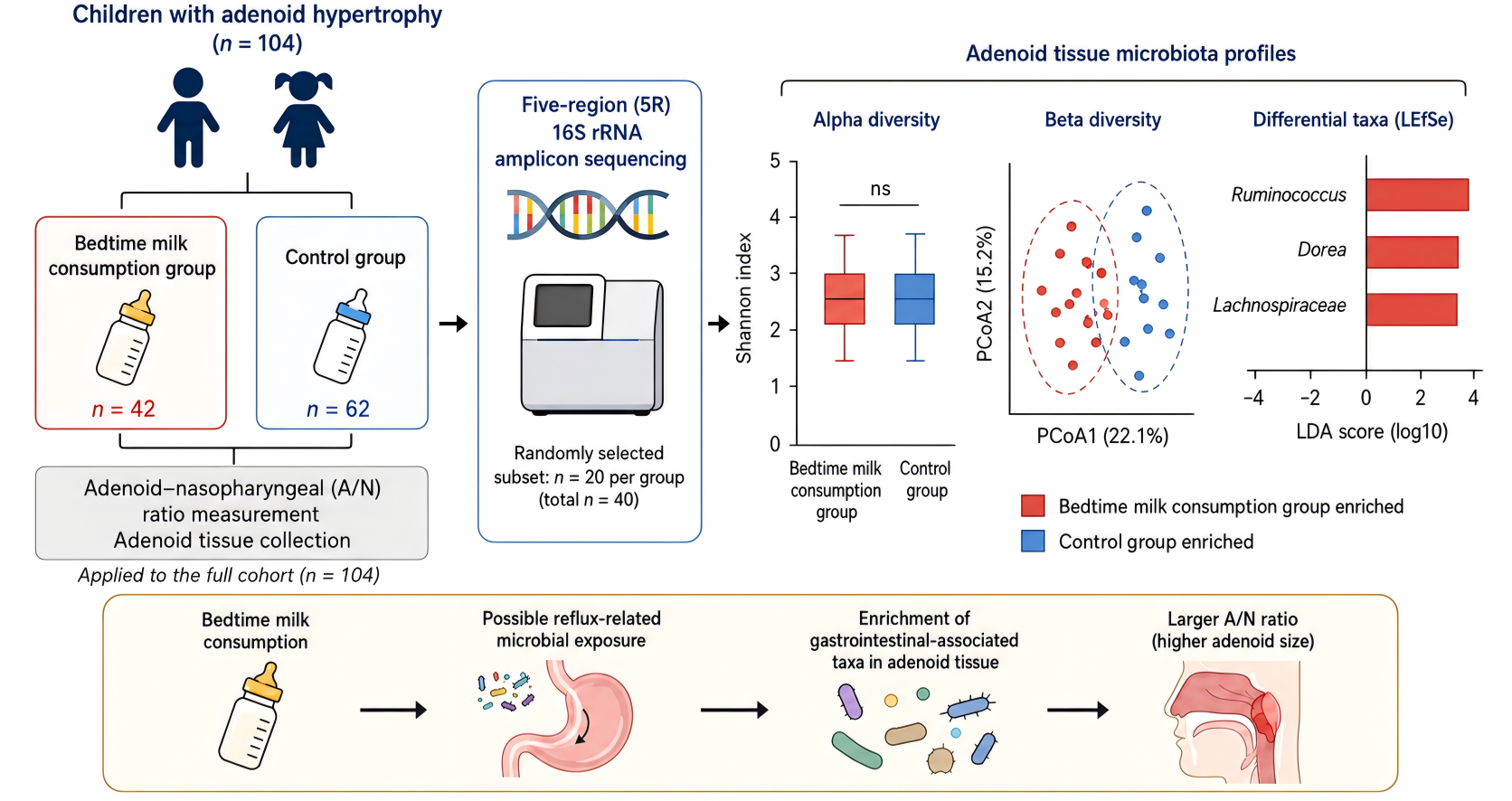
Background: Adenoid hypertrophy is common in children. This study investigated the association of bedtime milk consumption with adenoid size and tissue microbiota.
Methods: A total of 104 children undergoing surgery for adenoid hypertrophy were grouped by bedtime milk consumption. The adenoid-nasopharyngeal ratio was measured, and tissue samples from 20 participants in each group underwent five-region 16S rRNA sequencing.
Results: The average A/N ratio was higher in the bedtime milk group than in controls. Total microbial diversity and dominant bacterial distributions did not differ significantly, but Ruminococcus, Dorea, and Lachnospiraceae were enriched in the bedtime milk group.
Conclusions: Bedtime milk consumption was associated with larger adenoid size and altered tissue microbiota profiles. The findings suggest a possible reflux-related microbial exposure that requires further validation.
Keywords: milk drinking, adenoid hypertrophy, gastroesophageal reflux, microorganisms
Introduction
Adenoids, also known as pharyngeal tonsils, are located between the pharyngeal recesses on both sides at the junction of the roof and posterior wall of the nasopharynx. Adenoids are part of Waldeyer's ring and play an important role in immune system development. Adenoids are also the first organ of the immune system in contact with respiratory tract and digestive tract antigens and serve as a collective defense barrier against infection. Studies have shown that adenoids perform the earliest immune function in nasal-associated lymphoid tissue (NALT). Immune activity is strongest in the adenoids at 4-10 years of age in humans and begins to regress after puberty [1].
The number of children diagnosed with adenoid hypertrophy (AH) is increasing annually [2]. AH in children is typically accompanied by a series of clinical manifestations, such as nasal obstruction, oral breathing, snoring, hearing impairment, and changes in the structure and appearance of the craniofacial region [3]. Although these symptoms can be treated surgically, surgery has a psychological impact on children and parents and potentially results in complications, such as bleeding. These negative effects of surgical treatment have prompted doctors to investigate alternative nonsurgical means of treatment. However, the development of new diagnostic and treatment strategies has been challenging. One recent study indicated that drug therapy does not reduce the surgical rate in AH children. Nonsurgical treatment methods, such as the use of leukotriene receptor antagonists, nasal steroids, and/or oral antihistamines, are not directly linked to a decrease in the number of surgeries performed [4]. Although the causes of AH in children are incompletely understood, existing research suggests that AH may be related to infections, immune dysregulation, environmental exposure, hormonal factors, and genetic susceptibility [5–7]. Adenoid tissue is exposed to both inhaled and swallowed microorganisms and may therefore reflect microbial signals from the upper airway and digestive tract. Recent microbiome studies have suggested that AH is associated with altered local or intestinal microbial communities, but the relationship between daily feeding habits, reflux-related exposure, and adenoid tissue microbiota remains poorly understood. Because adenoid tissue contains relatively low microbial biomass, careful contamination control and negative controls are essential for reliable microbial profiling.
In clinical practice, bedtime milk consumption is common in children and may be associated with AH.. Therefore, we divided children with AH who underwent surgical treatment into groups according to bedtime milk consumption, measured the adenoid-nasopharyngeal (A/N) ratio, and performed five-region (5R) 16S rRNA amplicon sequencing on adenoid tissue samples to explore the association between bedtime milk consumption, adenoid size, and local microbial profiles.
Methods
Participant recruitment
The participants in this study comprised 104 children who underwent surgical treatment for AH at the Department of Otolaryngology-Head and Neck Surgery of the Affiliated Bozhou Hospital of Anhui Medical University between March 2023 and January 2024. The participants included 60 males and 44 females aged 3-10 (6.38 ± 1.81) years. All participants (legal guardians) provided written consent (provided by legal guardians and children over 7 years old) for their participation. The diagnosis of AH was achieved through nasal endoscopy and nasopharynx imaging. The enrolled children with AH had over 75% obstruction in their nasal airways, as confirmed by nasal endoscopy. Imaging was also performed to confirm an adenoid-nasopharyngeal (A/N) ratio greater than or equal to 0.7.
CT examination and measurement of the A/N ratio
Imaging was performed on the nasopharynx with the participants remaining calm and adopting a supine position. Computer software was used for data acquisition and analysis. In the A/N ratio, the adenoid size A is the vertical distance from the outermost point of convexity of the adenoid shadow to the tangent of the basiocciput, and the nasopharyngeal size N is the distance between the anterior edge of the sphenobasioccipital synchondrosis and the posterior edge of the hard palate. The A/N ratio was obtained by dividing two of the measured values. All measurements were obtained by the imaging department using professional software.
Inclusion, exclusion, and grouping criteria
Enrolled children with AH were surveyed on their bedtime milk consumption behavior. Children who consumed > 100 ml of milk 1 h before bedtime for more than 5 days per week were assigned to the experimental group. Children who did not consume milk before bedtime or consumed any other type of food 1 h before bedtime were grouped into the control group. The exclusion criteria were as follows: a) the presence of other potential diseases, such as cancer, gastrointestinal diseases, or inflammatory diseases; b) the use of antibiotics and probiotics in the month before the start of the study; c) surgical treatment within three months before the study; and d) incomplete clinical information.
Of the 104 patients selected for the study, 42 were grouped into the bedtime milk consumption group, including 24 males and 18 females aged 3-10 (6.19 ± 1.95) years. The control group comprised 62 patients, including 36 males and 26 females aged 3-9 (6.50 ± 1.72) years. The final grouping was 42 children in the bedtime milk consumption group and 62 children in the control group.
Sample collection and sequencing
The clinical symptoms of the children with AH were observed and recorded. Fresh tissue samples were collected from every child during surgery and stored at −80°C for subsequent experimental use. The following procedures were adopted: the nasopharynx of the children with AH was rinsed with saline water three times before adenoidectomy, and fresh adenoid tissue samples were collected during surgery. The samples were mung bean-sized with a length, width, and height of ≤0.5 cm. The samples were immediately washed with 1× PBS or saline prepared with sterile, nuclease-free water precooled to 2-6°C to remove any blood or stains from their surface. The samples were then dried with sterile and lint-free paper towels and quickly placed in precooled, sterile, and nuclease-free cryogenic vials with threaded caps that can withstand temperatures of −192°C. The vials were frozen in liquid nitrogen for 1 h and stored at −80°C for later use. Repeated freeze‒thaw cycles were avoided before DNA extraction. Due to the low bacterial content in the tissue samples and the difficulty in avoiding external bacterial contamination during sampling, negative control samples were collected along with the sequencing samples. More specifically, contaminating bacteria in the sampling environment were collected in a 1.5 mL sterile centrifuge tube. The tube was opened at the beginning of collection and closed at the end. Five negative control samples were collected to ensure the accurate identification of contaminating bacteria.
5R 16S rRNA amplicon sequencing
5R 16S rRNA amplicon sequencing, 20 children were randomly selected from the bedtime milk consumption group and 20 children were randomly selected from the control group. In this study, 5R refers to the parallel amplification and sequencing of five 16S rRNA amplicon regions spanning the following hypervariable segments: V2, V3, V4, V6-V7, and V8-V9. This approach differs from conventional single-region 16S rRNA sequencing, which usually relies on one amplicon, such as V3-V4 or V4, to profile microbial communities. By integrating sequence information from several non-contiguous 16S regions, the 5R approach provides broader taxonomic information for microbial community profiling. The short multiple regions framework (SMURF) [8] was then used to integrate reads from the five amplified regions and perform taxonomic identification of bacteria. Due to the low microorganism content in the tissue samples collected, any microorganisms from the environment introduced during sample collection, DNA extraction, and PCR could cause significant interference in the identification of microorganisms already present in the sample. Hence, a filtration step to remove contaminating microorganisms (negative controls) introduced from the above sources was performed [9]. Finally, bacterial data collected after the filtration step were used to analyze the intra- and intergroup bacterial diversity and perform comparisons among the bacterial communities. 5R 16S rRNA amplicon sequencing was performed by Hangzhou Lianchuan Biotechnology Co., Ltd.
Comparison of bacterial communities
Five-region 16S rRNA sequencing data were pooled and analyzed with SMURF to identify the bacterial communities in the sample and calculate the relative abundance. Specifically, SMURF uses the expectation-maximization algorithm to compile the sequencing data from multiple short, amplified regions and reconstruct them to find the most likely set of 16S sequences. Bacterial taxonomy identification was then performed, and the relative abundance was calculated. The optimized Greengenes (May 2013 version) database was used. To mitigate the impact of low-abundance noise on subsequent analysis, samples with a total number of reads < 1,000 (including negative controls) and bacterial data with a relative abundance of < 10−4 were removed. The number of reads was normalized for each sample. Bacterial contaminants introduced during sampling and experimentation were identified according to the prevalence of bacterial communities in the negative controls. In this study, a threshold of 50% prevalence was set for identifying bacterial contaminants. The bacterial communities remaining in the sample after removal of the contaminating bacteria were considered those present in adenoid tissue. Analyses of alpha diversity, beta diversity, and dominant bacterial communities and comparisons of bacterial communities were subsequently conducted using these data.
Statistical analysis
For the comparison of the A/N ratio between groups, the mean difference and 95% confidence interval (CI) were calculated using Welch's method and reported to improve statistical transparency.
Results
Essential clinical traits of participants with AH
A total of 104 children with AH fulfilled the inclusion and exclusion criteria described in the Materials and Methods section. Forty-two of them (40.4%) who regularly consumed milk 1 h before bedtime were included in the bedtime milk consumption group, including 24 males and 18 females aged 3-10 (6.19 ± 1.95) years. Sixty-two children (59.6%) who did not regularly consume milk before bedtime, including 36 males and 26 females aged 3-9 years (6.50 ± 1.72), were included in the control group. No significant differences in sex, age, body mass index (BMI), or disease onset time were observed between the two groups (Table 1).
Table 1. Clinical characteristics of children with adenoidal hypertrophy (AH).
| Clinical characteristic | Bedtime milk consumption group (n = 42) | Control group (n = 62) | t/χ² | P |
|---|---|---|---|---|
| Gender (male/female) | 24/18 | 36/26 | 0.009 | 0.926 |
| Age (years, mean ± SD) | 6.19 ± 1.95 | 6.50 ± 1.72 | 0.853 | 0.395 |
| BMI (kg*m−2, mean ± SD) | 16.50 ± 2.82 | 17.21 ± 3.47 | 1.147 | 0.254 |
| Disease onset time (years, mean ± SD) | 1.65 ± 1.36 | 1.42 ± 0.90 | −1.028 | 0.307 |
The A/N ratio was higher in children with bedtime milk consumption behavior
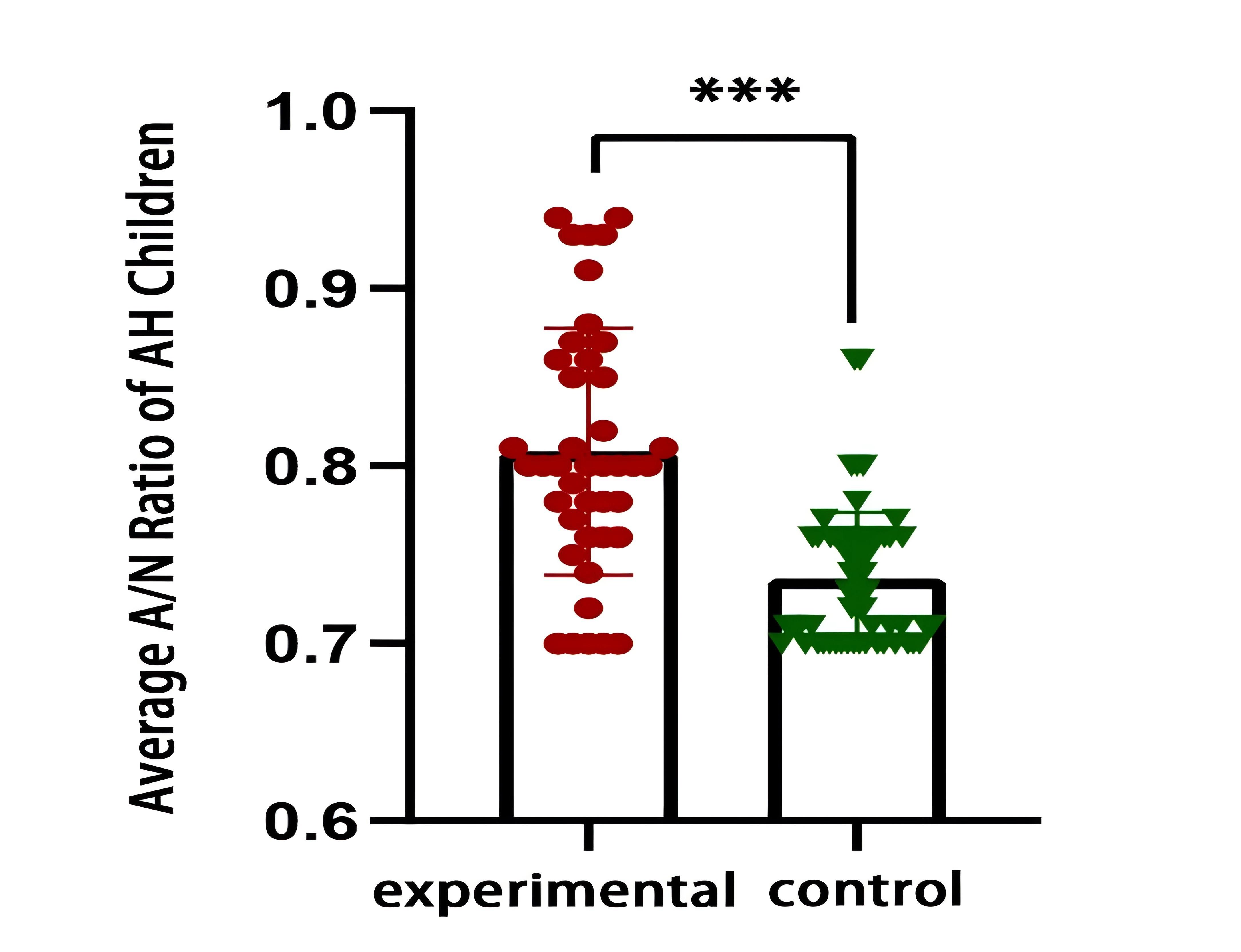
A higher average A/N ratio (0.808 ± 0.070) was observed in children who consumed milk before bedtime, whereas those who did not had an average A/N ratio of 0.736 ± 0.037. The mean difference between groups was 0.072 (95% CI, 0.048 to 0.096), and the intergroup difference was significant (P < 0.001) (Figure 1).
Figure 1. Comparison of the A/N ratio between the bedtime milk consumption group and the control group. Children in the bedtime milk consumption group had a higher average A/N ratio than those in the control group. The mean difference was 0.072 (95% CI, 0.048 to 0.096). P < 0.001.
Comparison of total bacterial diversity in adenoid tissue between children with AH with and without bedtime milk drinking behavior
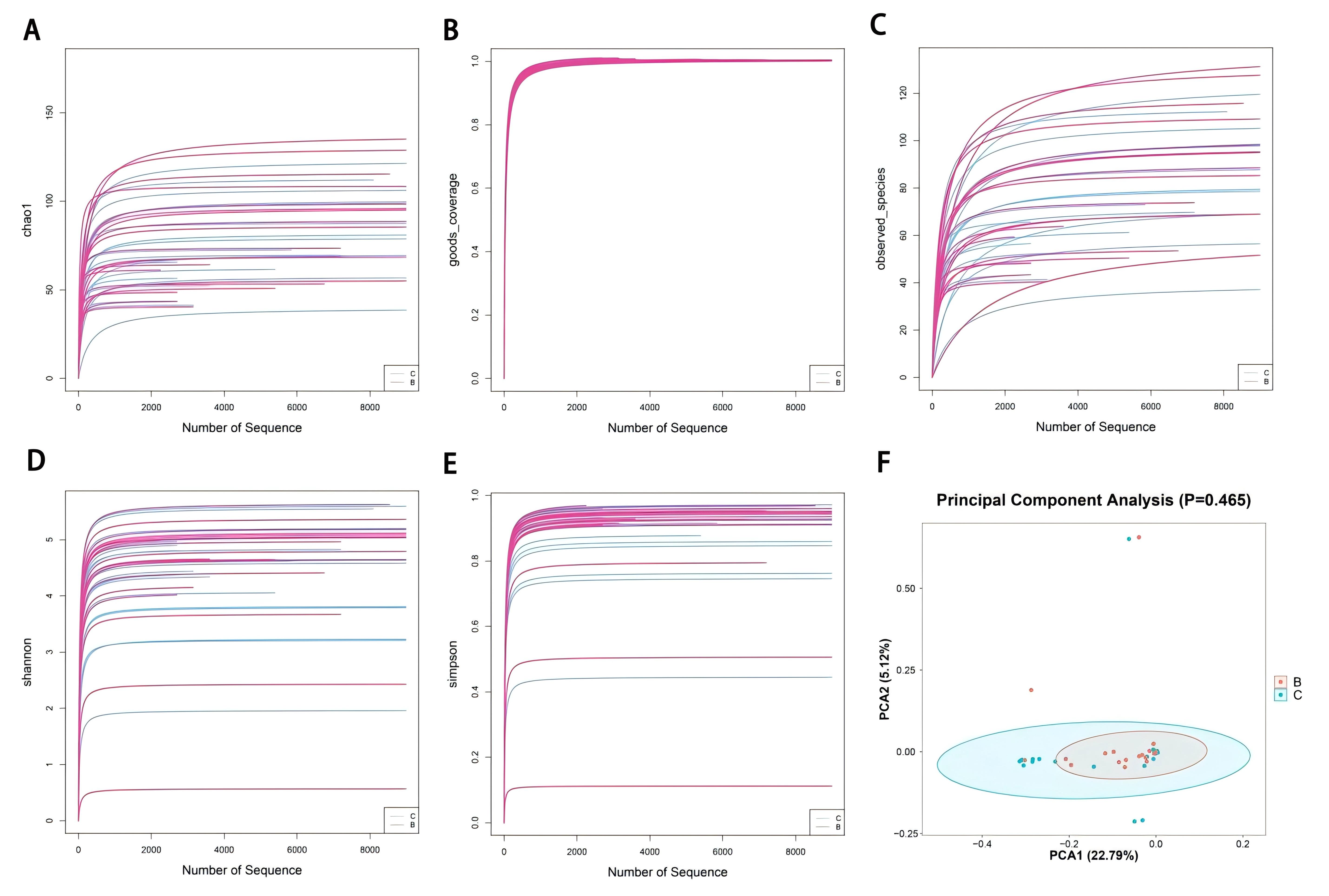
Alpha diversity is an indicator of diversity within a particular environment or ecosystem and is primarily used to reflect species richness and evenness and sequencing depth. Various alpha diversity metrics, such as the Chao1, Good’s coverage, observed species, Shannon, and Simpson indices (Figure 2A-E), were used to quantify species richness and evenness in this study. Beta diversity measures the species diversity between different ecological communities. Together, alpha and beta diversity constitute the total species diversity or biological heterogeneity of a certain ecological community. Analysis of the data obtained from the two groups of children with AH revealed no significant difference in the total bacterial diversity in their adenoid tissue regardless of bedtime milk consumption behaviors.
Figure 2. Characteristics of the total bacterial diversity in the adenoid tissue of the bedtime milk consumption group and the control group. (A-E) Alpha diversity indices. (F) Principal component analysis showing intragroup similarity and intergroup differences (P = 0.465).
Analysis of dominant bacterial communities in the adenoid tissue of AH children in the experimental and control groups
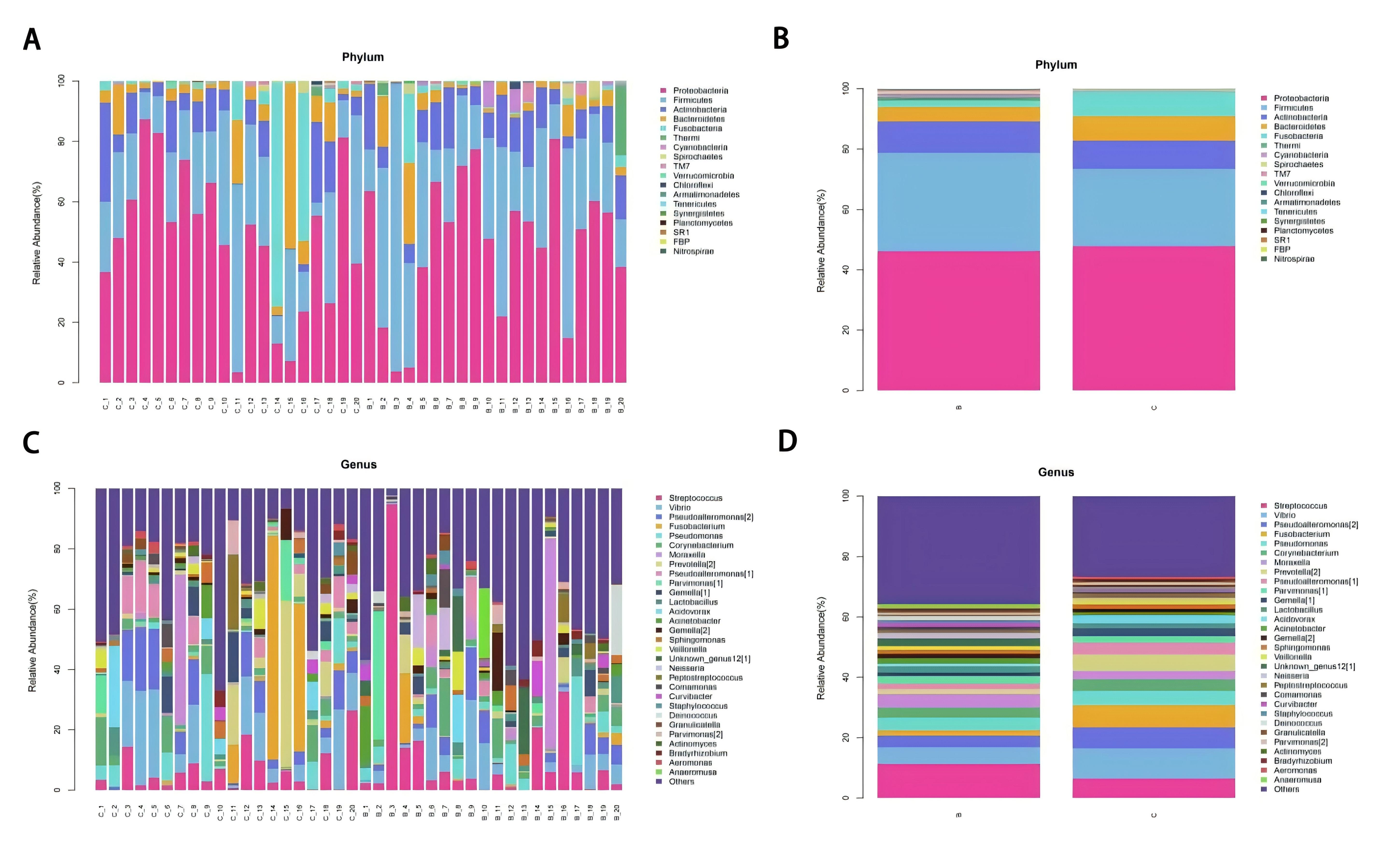
The distribution of bacterial communities in the adenoid tissue of the two groups of children with AH was analyzed at the domain, phylum, class, order, family, genus, and species levels. Figure 3 shows the results obtained at the phylum and genus levels. Figure 3A-B shows the overall distribution of bacteria in the adenoid tissue of the two groups of children at the phylum level and the intergroup comparisons. At the phylum level, Proteobacteria, Firmicutes, Actinobacteria, Bacteroidetes, and Fusobacteria were the dominant bacteria in the adenoid tissue of both groups. No significant difference in the distribution of dominant bacterial communities was observed between the experimental and control groups. Figure 3C-D shows the total bacterial distribution in the adenoid tissue of the two groups at the genus and intergroup levels. At the genus level, Streptococcus, Vibrio, Pseudoalteromonas, Fusobacterium, Pseudomonas, and Corynebacterium were the dominant bacteria in the adenoid tissue of both groups. No significant difference was detected in the distribution of the dominant bacterial communities between the experimental and control groups.
Figure 3. Analysis of the dominant bacterial communities in the adenoid tissue of the bedtime milk consumption group and the control group. (A, C) Average composition and relative abundance at phylum and genus levels. (B, D) Between-group comparisons.
Comparison of bacterial communities in the adenoid tissue of the two groups of children with AH
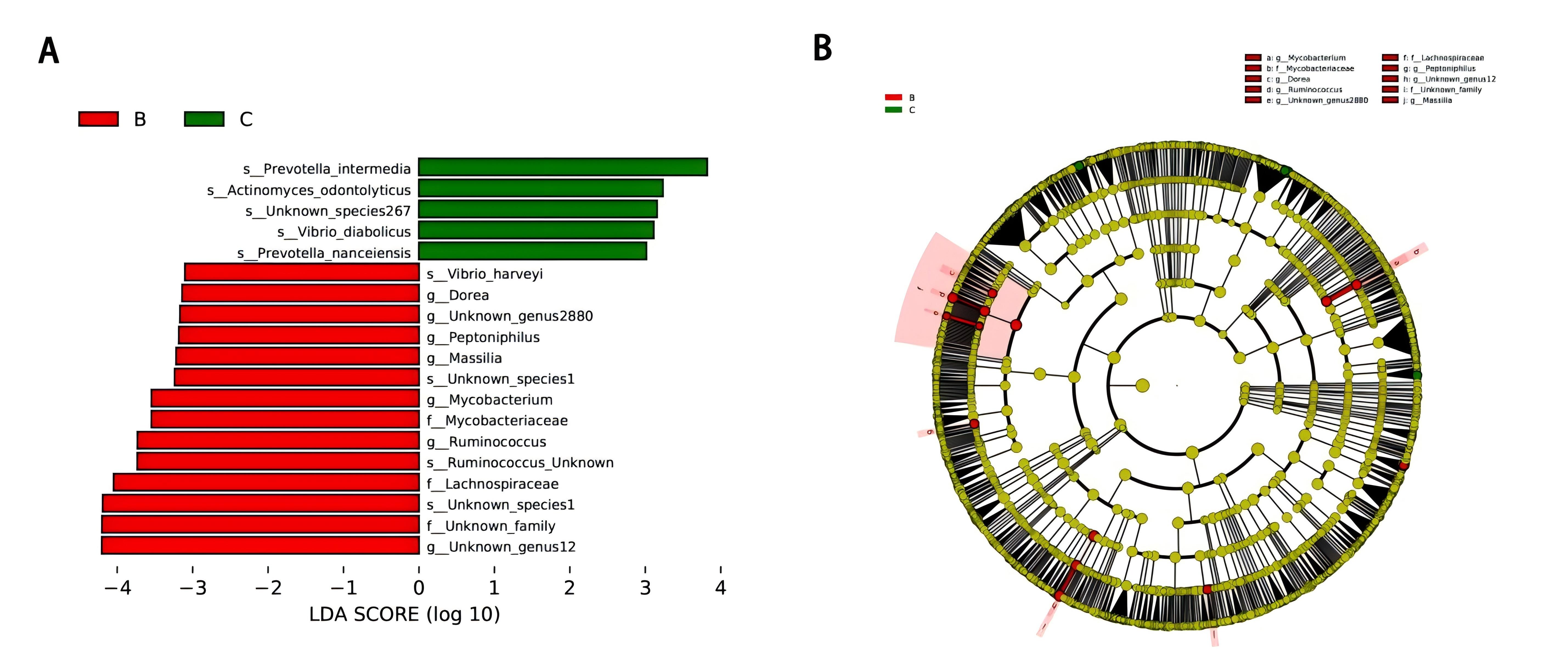
Linear discriminant analysis effect size (LEfSe) was used to analyze the differences in the bacterial communities between the two groups. The threshold value was set at LDA > 3 and P < 0.05. Compared to those in the control group, the adenoid tissue of children with AH who consumed bedtime milk was enriched in Ruminococcus, Mycobacterium, Dorea (a member of the Lachnospiraceae family), Lachnospiraceae, Peptidophilus, and Massilia (Figure 4). Ruminococcus, Dorea, and Lachnospiraceae commonly colonize the gastrointestinal tract.
Figure 4. Comparison of bacterial communities in the adenoid tissue of the bedtime milk consumption group and the control group. LEfSe analysis identified differential taxa. Ruminococcus, Dorea, and Lachnospiraceae were enriched in the bedtime milk consumption group. LDA > 3, P < 0.05.
Discussion
In recent years, there has been a steady increase in cases of AH in children [10]. Although surgical treatment has achieved good results, the cause of this disease is still unclear [10]. The increase in the number of children receiving surgery has prompted doctors to look into the cause and pathogenesis of AH and new diagnosis and treatment methods. Studies have shown that changes in microbial community structure could be related to various diseases in the human body [11–12], and scientists have previously investigated the relationship between microorganisms and AH [13–14]. In this study, 5R 16S rRNA amplicon sequencing was performed on adenoid tissue from children with AH. The dominant bacterial communities in adenoid tissue at the phylum level were Proteobacteria, Firmicutes, Actinobacteria, Bacteroidetes, and Fusobacteria. This result is consistent with the distribution of nasopharyngeal bacterial communities identified by Bogaert et al. [14]. At the genus level, Streptococcus, Vibrio, Pseudoalteromonas, Fusobacterium, Pseudomonas, and Corynebacterium were the dominant bacterial communities in adenoid tissue. Previous microbial analysis of adenoid tissue in children by Brook et al. also suggested a similar bacterial distribution [15]. The unique microbiology of AH and the interactions among various organisms revealed in this study provide new directions for antimicrobial therapy for AH.
In clinical practice, bedtime milk consumption is common among children and may be associated with AH. In this study, children in the bedtime milk consumption group had a higher average A/N ratio than those in the control group (0.808 ± 0.070 vs. 0.736 ± 0.037; mean difference, 0.072; 95% CI, 0.048 to 0.096; P < 0.001), suggesting an association between bedtime milk consumption and larger adenoid size. 5R 16S rRNA amplicon sequencing further showed that adenoid tissue from children in the bedtime milk consumption group was enriched in Ruminococcus, Mycobacterium, Dorea, Lachnospiraceae, Peptoniphilus, and Massilia. Among these taxa, Ruminococcus, Dorea, and Lachnospiraceae are commonly associated with the gastrointestinal tract [16–18]. Their enrichment in adenoid tissue may therefore reflect reflux-related microbial exposure, microbial translocation along the aerodigestive tract, or broader host–microbiome interactions involving mucosal immunity, diet, and local inflammatory status.
Previous studies have reported an association between GERD and AH. Niu et al. [19] reported a significant correlation between AH and GERD, and another study suggested that upper airway obstruction may be associated with increased GERD occurrence through changes in intrathoracic pressure [20]. Crapko et al. [21] also reported possible reflux-related inflammatory responses in adenoid tissue. Consistent with these findings, our results showed that children in the bedtime milk consumption group had larger adenoids and a higher abundance of gastrointestinal-associated taxa in adenoid tissue. However, these observations do not establish causality, and reflux involvement can only be inferred indirectly. This study was limited by its single-center observational design, the relatively small number of sequenced samples, the absence of direct reflux-related assessments such as pH/impedance monitoring or pepsin detection, potential unmeasured dietary confounders, and the lack of longitudinal follow-up.
In summary, bedtime milk consumption was associated with larger adenoid size and altered adenoid tissue microbiota profiles in children with AH. The enrichment of gastrointestinal-associated taxa may be linked to reflux-related microbial exposure; however, direct reflux-related examinations were not performed, and further prospective studies incorporating pH/impedance monitoring or pepsin detection are needed to clarify this association.
Abbreviations
5R: Five-region; 16S rRNA: 16S ribosomal RNA; A/N: Adenoid-nasopharyngeal ratio; AH: Adenoid hypertrophy; BMI: Body Mass Index; CI: Confidence Interval; CT: Computed Tomography; DNA: Deoxyribonucleic Acid; GERD: Gastroesophageal Reflux Disease; GSA: Genome Sequence Archive; LDA: Linear Discriminant Analysis; LEfSe: Linear Discriminant Analysis Effect Size; NALT: Nasal-associated Lymphoid Tissue; PBS: Phosphate-buffered Saline; PCA: Principal Component Analysis; PCR: Polymerase Chain Reaction; SD: Standard Deviation; SMURF: Short Multiple Regions Framework.
Declarations
Author Contributions
W.C. and L.W. contributed equally to this work. W.C. and L.W. led conceptualization, methodology, formal analysis, data interpretation, and drafting. W.C. led supervision, project administration, revision, and final approval. W.W., C.F., M.H., J.J., and M.Z. contributed to investigation, data curation, laboratory work, sample collection, and manuscript review. All authors approved the final manuscript.
Data availability
The raw sequence data have been deposited in the Genome Sequence Archive (GSA: CRA017958) and can be accessed at https://bigd.big.ac.cn/gsa/browse/CRA017958.
Funding
This work was supported by the Key Scientific Research Project and Development Plan of Bozhou (bzzc2023058).
Ethics approval and consent to participate
The study was performed in accordance with relevant guidelines and regulations and the principles of the Declaration of Helsinki. Ethical approval was obtained from the Medical Ethics Committee of Bozhou People’s Hospital. Informed consent was obtained from the parents or legal guardians, and assent was obtained from children aged 7 years or older.
Consent for publication
All authors have agreed to publish.
Competing Interests
The authors declare no potential conflicts of interest.
Clinical trial number
Not applicable.
References
[1] Nave, H., A. Gebert, and R. Pabst. Morphology and Immunology of the Human Palatine Tonsil. Anatomy and Embryology 204 (5): 367–373. https://doi.org/10.1007/s004290100210
[2] Pereira, Lara, John Monyror, Fabiana T. Almeida, Fernanda R. Almeida, and Eliete Guerra. Prevalence of Adenoid Hypertrophy: A Systematic Review and Meta-Analysis. Sleep Medicine Reviews 38: 101–112. https://doi.org/10.1016/j.smrv.2017.06.001
[3] Niedzielski, Artur, Lechosław Paweł Chmielik, Grażyna Mielnik-Niedzielska, Anna Kasprzyk, and Joanna Bogusławska. Adenoid Hypertrophy in Children: A Narrative Review of Pathogenesis and Clinical Relevance. BMJ Paediatrics Open 7 (1): e001710. https://doi.org/10.1136/bmjpo-2022-001710
[4] Tamir, S. O., Y. Schwarz, I. Hazan, et al. Medical Treatment Does Not Reduce Surgery Rates in Children with Adenoid Hypertrophy. International Journal of Pediatric Otorhinolaryngology 176: 111836. https://doi.org/10.1016/j.ijporl.2023.111836
[5] Ma, Y., L. Xie, and W. Wu. The Effects of Adenoid Hypertrophy and Oral Breathing on Maxillofacial Development: A Review of the Literature. Journal of Clinical Pediatric Dentistry 48 (1): 1–6.
[6] Hua, H. L., Y. Q. Deng, H. Huang, et al. Inflammatory Endotypes of Adenoidal Hypertrophy Based on a Cluster Analysis of Biomarkers. International Immunopharmacology 127: 111318. https://doi.org/10.1016/j.intimp.2023.111318
[7] Liu, W., H. Jiang, X. Liu, et al. Altered Intestinal Microbiota Enhances Adenoid Hypertrophy by Disrupting the Immune Balance. Frontiers in Immunology 14: 1277351. https://doi.org/10.3389/fimmu.2023.1277351
[8] Fuks, G., M. Elgart, A. Amir, et al. Combining 16S rRNA Gene Variable Regions Enables High-Resolution Microbial Community Profiling. Microbiome 6 (1): 17. https://doi.org/10.1186/s40168-017-0396-x
[9] Nejman, D., I. Livyatan, G. Fuks, et al. The Human Tumor Microbiome Is Composed of Tumor Type-Specific Intracellular Bacteria. Science 368 (6494): 973–980. https://doi.org/10.1126/science.aay9189
[10] Bilgili, A. M., H. Ö. Durmaz, and M. Dilber. Efficacy of Topical Azelastine and Fluticasone Dipropionate Combination in Children with Adenoid Hypertrophy. Ear, Nose & Throat Journal 102 (1): 28–34.
[11] Zhou, B., Y. Yuan, S. Zhang, et al. Intestinal Flora and Disease Mutually Shape the Regional Immune System in the Intestinal Tract. Frontiers in Immunology 11: 575. https://doi.org/10.3389/fimmu.2020.00575
[12] Zuo, L., L. He, A. Huang, et al. Risk Factors and Antibiotic Sensitivity of Aerobic Bacteria in Chinese Children with Adenoid Hypertrophy. BMC Pediatrics 22: 553..
[13] Ren, T., D. U. Glatt, T. N. Nguyen, et al. 16S rRNA Survey Revealed Complex Bacterial Communities and Evidence of Bacterial Interference on Human Adenoids. Environmental Microbiology 15 (2): 535–547.
[14] Bogaert, D., B. Keijser, S. Huse, et al. Variability and Diversity of Nasopharyngeal Microbiota in Children: A Metagenomic Analysis. PLoS ONE 6 (2): e17035. https://doi.org/10.1371/journal.pone.0017035
[15] Brook, I., and A. E. Gober. In Vitro Bacterial Interference in the Nasopharynx of Otitis Media-Prone and Non-Otitis Media-Prone Children. Archives of Otolaryngology–Head & Neck Surgery 126 (8): 1011–1013. https://doi.org/10.1001/archotol.126.8.1011
[16] Shi, Y. C., S. T. Cai, Y. P. Tian, et al. Effects of Proton Pump Inhibitors on the Gastrointestinal Microbiota in Gastroesophageal Reflux Disease. Genomics, Proteomics & Bioinformatics 17 (1): 52–63. https://doi.org/10.1016/j.gpb.2018.12.004
[17] Zhou, J., P. Shrestha, Z. Qiu, et al. Distinct Microbiota Dysbiosis in Patients with Non-Erosive Reflux Disease and Esophageal Adenocarcinoma. Journal of Clinical Medicine 9 (7): 2162. https://doi.org/10.3390/jcm9072162
[18] Zhang, X., D. Yu, D. Wu, et al. Tissue-Resident Lachnospiraceae Family Bacteria Protect against Colorectal Carcinogenesis by Promoting Tumor Immune Surveillance. Cell Host & Microbe 31 (3): 418–432.e8..
[19] Niu, Xun, Zeng-Hong Wu, Xi-Yue Xiao, and Xiong Chen. The Relationship between Adenoid Hypertrophy and Gastroesophageal Reflux Disease: A Meta-Analysis. Medicine 97 (41): e12540. https://doi.org/10.1097/MD.0000000000012540
[20] Sagar, M., P. Sagar, S. K. Kabra, et al. The Concatenation of Association between Gastroesophageal Reflux and Obstructive Adenotonsillar Hypertrophy. International Journal of Pediatric Otorhinolaryngology 139: 110439. https://doi.org/10.1016/j.ijporl.2020.110439
[21] Crapko, M., J. E. Kerschner, M. Syring, et al. Role of Extra-Esophageal Reflux in Chronic Otitis Media with Effusion. The Laryngoscope 117 (8): 1419–1423..
 Figures
 References
 Peer
 Information
Figure 1. Comparison of the A/N ratio between the bedtime milk consumption group and the control group. Children in the bedtime milk consumption group had a higher average A/N ratio than those in the control group. The mean difference was 0.072 (95% CI, 0.048 to 0.096). *P < 0.001.
Figure 2. Characteristics of the total bacterial diversity in the adenoid tissue of the bedtime milk consumption group and the control group. (A-E) Alpha diversity indices. (F) Principal component analysis showing intragroup similarity and intergroup differences (P = 0.465).
Figure 3. Analysis of the dominant bacterial communities in the adenoid tissue of the bedtime milk consumption group and the control group. (A, C) Average composition and relative abundance at phylum and genus levels. (B, D) Between-group comparisons.
Figure 4. Comparison of bacterial communities in the adenoid tissue of the bedtime milk consumption group and the control group. LEfSe analysis identified differential taxa. Ruminococcus, Dorea, and Lachnospiraceae were enriched in the bedtime milk consumption group. LDA > 3, P < 0.05.
[1] Nave, H., A. Gebert, and R. Pabst. Morphology and Immunology of the Human Palatine Tonsil. Anatomy and Embryology 204 (5): 367–373. https://doi.org/10.1007/s004290100210
[2] Pereira, Lara, John Monyror, Fabiana T. Almeida, Fernanda R. Almeida, and Eliete Guerra. Prevalence of Adenoid Hypertrophy: A Systematic Review and Meta-Analysis. Sleep Medicine Reviews 38: 101–112. https://doi.org/10.1016/j.smrv.2017.06.001
[3] Niedzielski, Artur, Lechosław Paweł Chmielik, Grażyna Mielnik-Niedzielska, Anna Kasprzyk, and Joanna Bogusławska. Adenoid Hypertrophy in Children: A Narrative Review of Pathogenesis and Clinical Relevance. BMJ Paediatrics Open 7 (1): e001710. https://doi.org/10.1136/bmjpo-2022-001710
[4] Tamir, S. O., Y. Schwarz, I. Hazan, et al. Medical Treatment Does Not Reduce Surgery Rates in Children with Adenoid Hypertrophy. International Journal of Pediatric Otorhinolaryngology 176: 111836. https://doi.org/10.1016/j.ijporl.2023.111836
[5] Ma, Y., L. Xie, and W. Wu. The Effects of Adenoid Hypertrophy and Oral Breathing on Maxillofacial Development: A Review of the Literature. Journal of Clinical Pediatric Dentistry 48 (1): 1–6.
[6] Hua, H. L., Y. Q. Deng, H. Huang, et al. Inflammatory Endotypes of Adenoidal Hypertrophy Based on a Cluster Analysis of Biomarkers. International Immunopharmacology 127: 111318. https://doi.org/10.1016/j.intimp.2023.111318
[7] Liu, W., H. Jiang, X. Liu, et al. Altered Intestinal Microbiota Enhances Adenoid Hypertrophy by Disrupting the Immune Balance. Frontiers in Immunology 14: 1277351. https://doi.org/10.3389/fimmu.2023.1277351
[8] Fuks, G., M. Elgart, A. Amir, et al. Combining 16S rRNA Gene Variable Regions Enables High-Resolution Microbial Community Profiling. Microbiome 6 (1): 17. https://doi.org/10.1186/s40168-017-0396-x
[9] Nejman, D., I. Livyatan, G. Fuks, et al. The Human Tumor Microbiome Is Composed of Tumor Type-Specific Intracellular Bacteria. Science 368 (6494): 973–980. https://doi.org/10.1126/science.aay9189
[10] Bilgili, A. M., H. Ö. Durmaz, and M. Dilber. Efficacy of Topical Azelastine and Fluticasone Dipropionate Combination in Children with Adenoid Hypertrophy. Ear, Nose & Throat Journal 102 (1): 28–34.
[11] Zhou, B., Y. Yuan, S. Zhang, et al. Intestinal Flora and Disease Mutually Shape the Regional Immune System in the Intestinal Tract. Frontiers in Immunology 11: 575. https://doi.org/10.3389/fimmu.2020.00575
[12] Zuo, L., L. He, A. Huang, et al. Risk Factors and Antibiotic Sensitivity of Aerobic Bacteria in Chinese Children with Adenoid Hypertrophy. BMC Pediatrics 22: 553..
[13] Ren, T., D. U. Glatt, T. N. Nguyen, et al. 16S rRNA Survey Revealed Complex Bacterial Communities and Evidence of Bacterial Interference on Human Adenoids. Environmental Microbiology 15 (2): 535–547.
[14] Bogaert, D., B. Keijser, S. Huse, et al. Variability and Diversity of Nasopharyngeal Microbiota in Children: A Metagenomic Analysis. PLoS ONE 6 (2): e17035. https://doi.org/10.1371/journal.pone.0017035
[15] Brook, I., and A. E. Gober. In Vitro Bacterial Interference in the Nasopharynx of Otitis Media-Prone and Non-Otitis Media-Prone Children. Archives of Otolaryngology–Head & Neck Surgery 126 (8): 1011–1013. https://doi.org/10.1001/archotol.126.8.1011
[16] Shi, Y. C., S. T. Cai, Y. P. Tian, et al. Effects of Proton Pump Inhibitors on the Gastrointestinal Microbiota in Gastroesophageal Reflux Disease. Genomics, Proteomics & Bioinformatics 17 (1): 52–63. https://doi.org/10.1016/j.gpb.2018.12.004
[17] Zhou, J., P. Shrestha, Z. Qiu, et al. Distinct Microbiota Dysbiosis in Patients with Non-Erosive Reflux Disease and Esophageal Adenocarcinoma. Journal of Clinical Medicine 9 (7): 2162. https://doi.org/10.3390/jcm9072162
[18] Zhang, X., D. Yu, D. Wu, et al. Tissue-Resident Lachnospiraceae Family Bacteria Protect against Colorectal Carcinogenesis by Promoting Tumor Immune Surveillance. Cell Host & Microbe 31 (3): 418–432.e8..
[19] Niu, Xun, Zeng-Hong Wu, Xi-Yue Xiao, and Xiong Chen. The Relationship between Adenoid Hypertrophy and Gastroesophageal Reflux Disease: A Meta-Analysis. Medicine 97 (41): e12540. https://doi.org/10.1097/MD.0000000000012540
[20] Sagar, M., P. Sagar, S. K. Kabra, et al. The Concatenation of Association between Gastroesophageal Reflux and Obstructive Adenotonsillar Hypertrophy. International Journal of Pediatric Otorhinolaryngology 139: 110439. https://doi.org/10.1016/j.ijporl.2020.110439
[21] Crapko, M., J. E. Kerschner, M. Syring, et al. Role of Extra-Esophageal Reflux in Chronic Otitis Media with Effusion. The Laryngoscope 117 (8): 1419–1423..
Peer-review Terminology
Identity transparency: Single-anonymous
Reviewer interacts with: Editor
Details
This is an open access article under the terms of the Creative Commons Attribution License(http://creativecommons.org/licenses/by/4.0/), which permits use, distribution and reproduction in any medium, provided the original work is properly cited.
Publication History
Received 2026-06-08
Accepted 2026-06-21
Published 2026-07-16