



Abstract
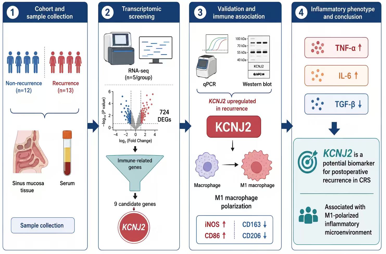
Background and objective: Chronic rhinosinusitis is a prevalent inflammatory disease with a relatively high postoperative recurrence rate. This study aimed to characterize transcriptomic features of recurrent disease and identify recurrence-associated biomarkers.
Methods: Twenty-five patients with chronic rhinosinusitis were enrolled, including 13 with postoperative recurrence and 12 without recurrence. Nasal mucosal tissues and serum samples were analyzed using RNA sequencing, RT-qPCR, Western blotting, and ELISA.
Results: KCNJ2 expression was significantly upregulated in the postoperative recurrence group and positively correlated with estimated M1 macrophage infiltration and inflammatory cytokines.
Conclusion: KCNJ2 may represent a candidate recurrence-associated biomarker, although prospective validation is required.
Keywords: Chronic rhinosinusitis; RNA-seq; Macrophages; KCNJ2; Recurrence
Introduction
CRS is a common upper respiratory tract disease, mainly caused by infections, allergies, or immune system abnormalities, and is characterized by chronic inflammation of the sinus mucosa [1–2]. The typical pathological changes of this disease include inflammatory cell infiltration, mucosal edema, glandular hyperplasia, and secretion retention [3–4]. Patients usually present with symptoms such as nasal congestion, rhinorrhea, hyposmia, and headache, which significantly impair their quality of life [5–6]. Epidemiological studies have shown that the prevalence of chronic sinusitis in China is approximately 8% and is increasing year by year [7]. Currently, the main treatment methods include nasal irrigation with normal saline and local steroid therapy [8–9]. If the therapeutic effect of drugs is poor, functional endoscopic sinus surgery (FESS) can be considered, which can effectively improve clinical symptoms [10]. However, studies have demonstrated that the postoperative recurrence rate remains relatively high [11], which not only increases the disease burden on patients but also imposes significant social and economic pressure. Therefore, identifying biomarkers related to postoperative recurrence is of great significance for in-depth understanding of the disease mechanism and improvement of prognosis management.
Transcriptomics, as an important tool for exploring disease pathogenesis, has been widely applied in the research of chronic sinusitis and its related disorders. For instance, Liu et al. identified four potential diagnostic biomarkers associated with nasal epithelial cell dysfunction by integrating multiple transcriptomic datasets of chronic sinusitis with nasal polyps (CRSwNP) [12]. Wang et al. combined single-cell sequencing with transcriptomic sequencing and found that dendritic cells are the key immune cell subset in CRSwNP; they further identified three hub genes, namely NR4A1, CLEC4G, and CD163, suggesting that these genes may serve as disease biomarkers and potential therapeutic targets [13]. Additionally, analysis based on transcriptomic data indicated that SLC17A4 was significantly downregulated in patients with CRS, and this finding was verified in clinical samples, rendering it a promising biomarker and therapeutic intervention target [14]. However, current studies focusing on analyzing the postoperative recurrence mechanism of CRS based on transcriptomic characteristics remain relatively limited.
The immune microenvironment plays a significant role in various inflammatory diseases. As key effector cells of the immune system, macrophages are crucial for regulating inflammation and maintaining the immune homeostasis of the nasal mucosa [15]. Under different microenvironmental stimuli, macrophages can differentiate into M1 and M2 macrophages. Upon activation, M1 macrophages secrete large amounts of pro-inflammatory factors, thereby promoting the inflammatory phenotype, while M2 macrophages are mainly involved in anti-inflammatory responses and tissue repair [16–17]. Studies have demonstrated that the infiltration of both M1 and M2 macrophages is significantly increased in patients with CRS. Hypoxia-inducible factor 1α (HIF1-α) can improve CRS by downregulating transferrin receptor (TFRC) expression and inhibiting M1 macrophage polarization. In patients with CRSwNP, the infiltration of M2 macrophages is more prominent [18–19], indicating the crucial role of macrophages in driving the progression of CRS. However, the mechanism underlying the role of macrophage polarization in the pathogenesis of CRS remains unclear. Therefore, it is essential to deeply understand the differentiation, phenotype, and function of macrophages in CRS.
This study aims to screen and identify key molecular markers closely associated with postoperative recurrence using transcriptome sequencing technology, followed by verification in clinical tissue samples. Based on these findings, it will further explore the intrinsic correlation between these markers and macrophage polarization in the immune microenvironment, thereby providing key scientific evidence for elucidating the pathological mechanism of postoperative recurrence, improving patient prognosis, and enhancing their quality of life.
Methods
Patient and Sample Collection
All participants provided written informed consent. The diagnosis of CRS was based on the criteria outlined in the European Position Paper on Rhinosinusitis and Nasal Polyps 2020 (EPOS 2020). The exclusion criteria are as follows: (1) Fungal sinusitis, allergic fungal sinusitis or malignant tumors; (2) Autoimmune diseases and other allergic or eosinophilic diseases; (3) Received immunotherapy, antibiotics, nasal/systemic steroids or anti-allergy drugs within 4 weeks before recruitment. A total of 25 patients with CRS were enrolled, including 12 patients without postoperative recurrence and 13 patients with recurrence. All participants provided written informed consent. Five samples from each group were randomly selected for RNA-seq. Initial RT-qPCR and Western blotting validation was performed using three biological samples per group, whereas expanded RT-qPCR, macrophage-marker analysis, and ELISA were performed using ten patients per group. The remaining samples were not included in specific assays because of insufficient sample volume or failure to meet assay-specific quality requirements. The RNA-seq samples were independent of the expanded validation cohort.
RNA-seq
Total RNA was extracted from human nasal mucosal tissue samples using TRIzol Reagent (Life Technologies, California, USA) according to the manufacturer's instructions. RNA concentration and purity were measured using a NanoDrop 2000 spectrophotometer (Thermo Fisher Scientific, Wilmington, DE, USA), and RNA integrity was assessed using the RNA Nano 6000 Assay Kit on an Agilent 2100 Bioanalyzer (Agilent Technologies, CA, USA). A total amount of 1 μg RNA per sample was used as input material for the RNA sample preparations. Sequencing libraries were generated using Hieff NGS Ultima Dual-mode mRNA Library Prep Kit for Illumina (Yeasen Biotechnology (Shanghai) Co., Ltd.) following manufacturer's recommendations and index codes were added to attribute sequences to each sample. Briefly, mRNA was purified from total RNA using poly-T oligo-attached magnetic beads. First strand cDNA was synthesized and second strand cDNA synthesis was subsequently performed. Remaining overhangs were converted into blunt ends via exonuclease/polymerase activities. After adenylation of 3' ends of DNA fragments, NEBNext Adaptor with hairpin loop structure were ligated to prepare for hybridization. The library fragments were purified with AMPure XP system (Beckman Coulter, Beverly, USA). Then 3 μl USER Enzyme (NEB, USA) was used with size-selected, adaptor-ligated cDNA at 37°C for 15 min followed by 5 min at 95°C before PCR. Then PCR was performed with Phusion High-Fidelity DNA polymerase, Universal PCR primers and Index (X) Primer. At last, PCR products were purified (AMPure XP system) and library quality was assessed on the Agilent Bioanalyzer 2100 system. The libraries were sequenced on an Illumina NovaSeq platform to generate 150 bp paired-end reads, according to the manufacturer's instructions.
Bioinformatics analysis
Differential expression analysis of two conditions/groups was performed using the DESeq2. DESeq2 provide statistical routines for determining differential expression in digital gene expression data using a model based on the negative binomial distribution. The resulting P values were adjusted using the Benjamini and Hochberg's approach for controlling the false discovery rate. Genes with an adjusted P-value < 0.01 & Fold Change ≥ 2 found by DESeq2 were assigned as differentially expressed.
Enrichment analysis
To identify candidate genes by crossing DEG and immune-related gene sets, The VennDiagram was drawn using the "Venn diagram" package. Enrichment analysis of these candidate genes was conducted using the ClusterProfiler software package (v3.18.1) in R, including the Gene Ontology (GO) and Kyoto Encyclopedia of Genes and Genomes (KEGG) pathways [20].
Immune infiltration analysis
Based on the transcriptome data, the immune infiltration was quantified using the "CIBERSORT" software package according to the CIBERSORT algorithm, which yielded the relative abundance of 22 immune cell types. The correlation between the target gene and these immune cells was then calculated via Spearman correlation analysis, and the lollipop plots was generated using the "ggplot2" package.
RT-qPCR
High-quality RNA was isolated from the sample to maintain both its integrity and purity. The extracted RNA was then converted into complementary DNA (cDNA) through reversed transcription using reverse transcriptase. Subsequently, targeted amplification of the cDNA was performed via PCR technology, utilizing appropriate primers and reaction conditions to acquire the desired fragment. The PCR process employed SYBR Premix Ex Taq™ (TaKaRa) and was carried out on the ABI StepOne™ Real-Time PCR System (Applied Biosystems, CA). The results were calculated using the 2-ΔΔCT method. GAPDH was used as the internal reference. The specific primers for qPCR are shown in Table 1.
Table 1. List of Primer Sequences.
| Primer | Sequences (5′-3′) |
|---|---|
| KCNJ2 F | TTCAGTCACAATGCCGTGATT |
| KCNJ2 R | GCTTTTCCGAAGATTGCCCA |
| KCNMB2 F | GAGGACCGAGCTATTCTCCTG |
| KCNMB2 R | TGTTTCCGTGATGGACGCATT |
| SLC4A4 F | TGATCGGGAGGCTTCTTCTCT |
| SLC4A4 R | GGACCGAAGGTTGGATTTCTTG |
| GNA15 F | CCAGGACCCCTATAAAGTGACC |
| GNA15 R | GCTGAATCGAGCAGGTGGAAT |
| APLNR F | CTCTGGACCGTGTTTCGGAG |
| APLNR R | GGTACGTGTAGGTAGCCCACA |
| EDN1 F | AGAGTGTGTCTACTTCTGCCA |
| EDN1 R | CTTCCAAGTCCATACGGAACAA |
Western Blotting
Sinus mucosal tissues were lysed in RIPA lysis buffer supplemented with 1 mM phenylmethylsulfonyl fluoride (PMSF; Bioteke Co., Wuxi, China). Equal amounts of protein were separated on 10% SDS-PAGE gels and transferred onto polyvinylidene fluoride (PVDF) membranes. The membranes were blocked at room temperature for 1 hour in TBST buffer containing 5% skimmed milk. Then, the membranes were incubated overnight at 4°C with anti-KCNJ2 (DF8117, Affinity, UK), and anti-GAPDH (60004-1-Ig, Proteintech, China). The next day, the membranes were incubated with horseradish peroxidase-conjugated goat anti-rabbit IgG secondary antibody (abs20040, absin, China) or goat anti-mouse horseradish peroxidase-conjugated secondary antibody (abs20039, absin) at room temperature for 1 hour. Then, the membranes were washed 3 times with TBST. The enhanced chemiluminescence (ECL) detection kit was used to detect protein bands and quantitative analysis was performed using ImageJ software.
ELISA
Blood samples were collected and homogenized at room temperature for 30 minutes. The samples were then centrifuged at 2000×g for 10 minutes at 4°C. The supernatant obtained after centrifugation was collected, aliquoted, and stored at temperatures below -20°C for subsequent analysis. Commercial ELISA kits were used to measure the serum concentrations of TNF-α (EH111776, BYabscience, China), IL-6 (EH110377, BYabscience), and TGF-β (EH1100532, BYabscience) following the manufacturer's instructions.
Statistical analysis
Data are presented as the mean ± standard deviation. Normality and homogeneity of variance were assessed using the Shapiro-Wilk test and Levene's test, respectively. Comparisons between two groups were performed using an unpaired two-tailed Student's t-test when these assumptions were met. Welch's t-test or the Mann-Whitney U test was used when appropriate. Categorical variables were compared using Fisher's exact test. Correlations were evaluated using Spearman's rank correlation analysis. All data points represent independent patients. Exact P values were reported where feasible, and P < 0.05 was considered statistically significant.
Result
Clinical characteristics of the subjects
A total of 25 patients were enrolled in this study, including 12 in the CRS non-recurrence group and 13 in the postoperative recurrence group. The baseline characteristics of the two groups are presented in Table 2. In the non-recurrence group, there were 7 males and 5 females, whereas the recurrence group consisted of 11 males and 2 females. There was no significant difference in sex distribution between the two groups (Fisher's exact test, P = 0.202). Regarding age, the non-recurrence group had a mean age of (39 ± 14) years, while the recurrence group had a mean age of (32 ± 12) years (P = 0.071). For body mass index (BMI), the non-recurrence group had an average of (25 ± 3) kg/m², compared with (28 ± 5) kg/m² in the recurrence group (P = 0.064). Statistical analysis revealed no statistically significant differences in age or BMI between (P > 0.05).
Table 2. Overall Patient Demographic Characteristics and Associated Clinical Features.
| Characteristic | Non-recurrence group (n = 12) | Recurrence group (n = 13) | P value |
|---|---|---|---|
| Male/female | 7/5 | 11/2 | / |
| Age (years) | 39±14 | 32±12 | 0.071 |
| BMI (kg/m²) | 25±3 | 28±5 | 0.064 |
Transcriptomic analysis reveals the specific transcriptome associated with the recurrence of CRS
To identify the biomarkers for the recurrence of chronic sinusitis after surgery, 5 samples were randomly selected from each of the two groups for transcriptome sequencing. The transcriptomic characteristics of the two groups of samples were shown in Figure 1. PCA revealed that the samples from the non-recurrence group and the recurrence group exhibited independent clustering trends, indicating significant differences in transcriptomic expression between the two groups (Figure 1A). Volcano plot analysis showed that a total of 724 differentially expressed genes (DEGs) were identified, among which 543 genes exhibited significantly upregulated expression and 181 genes exhibited significantly downregulated expression (Figure 1B-C). Subsequently, we performed GO enrichment analysis on the DEGs, and the results showed that these DEGs were mainly enriched in processes related to ciliary structural integrity, motor function, or ciliogenesis and ciliary structure and dysfunction are also one of the pathophysiological characteristics of chronic sinusitis (Figure 1D) [21]. Further KEGG pathway enrichment analysis revealed that the DEGs were mainly involved in immune inflammation-related pathways and the PPAR signaling pathway (Figure 1E), suggesting that they might participate in the recurrence process of sinusitis after surgery by regulating these pathways.
Figure 1. RNA-seq identified a distinctive gene profile in CRS patients between the Recurrence and Non-recurrence groups.
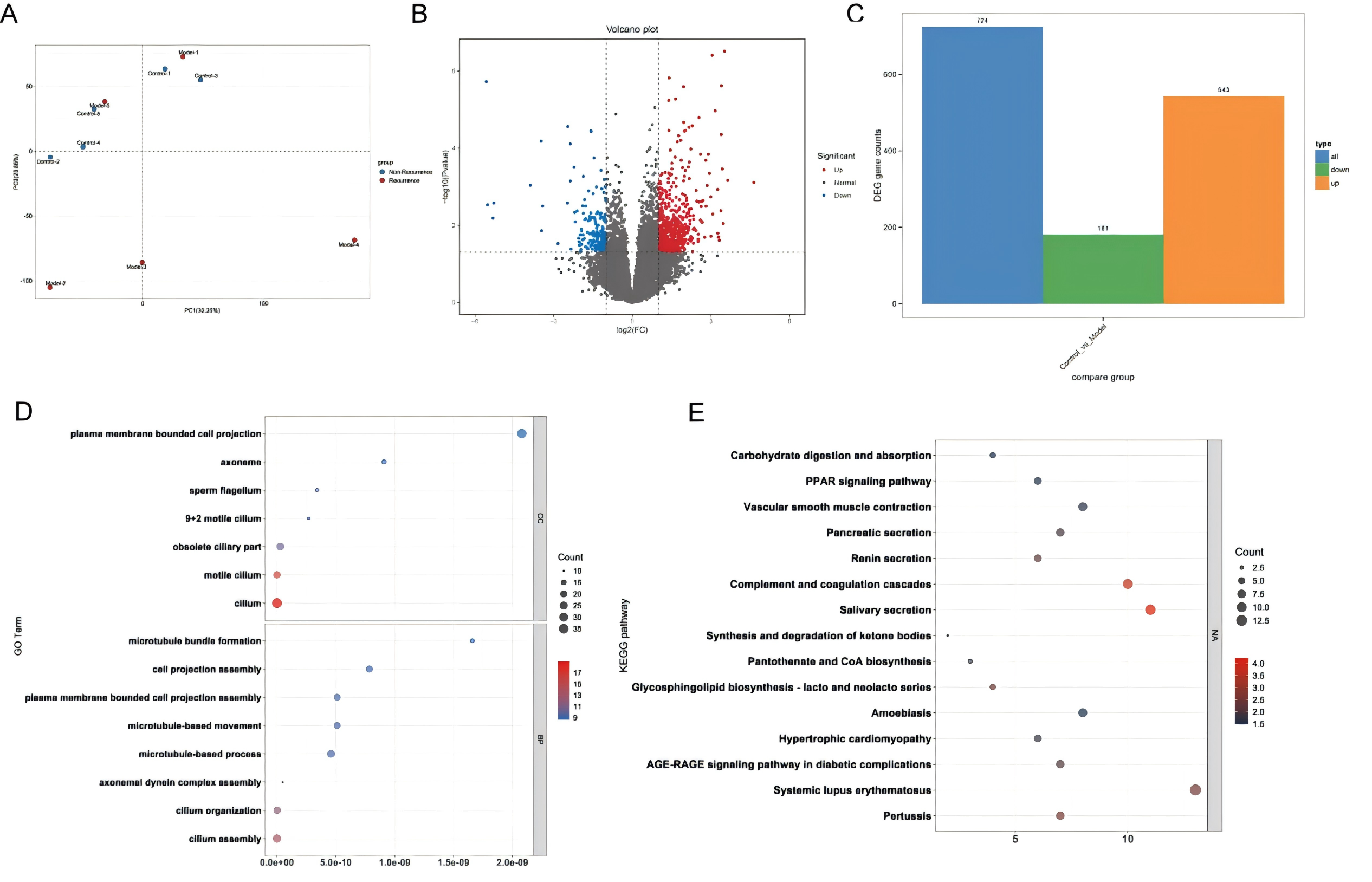
(A) PCA model of the transcriptomics data between the two groups. (B) Volcano plot showing genes upregulated and downregulated by RNA-seq. (C) Bar chart shows the number of DEGs between the two groups. (D) GO enrichment analysis. (E) KEGG enrichment analysis. Control, Non-recurrence; Model, Recurrence. n=5.
KCNJ2 was significantly elevated in the recurrence group and was associated with M1 macrophages
Given that the immune microenvironment plays an important role in CRS, we downloaded immune-related gene sets from the database and took the intersection with the DEGs obtained by RNA-seq. As shown in Figure 2A, 9 DEGs were finally obtained. The expression heatmap showed that EDN1 and APLNR were downregulated in the postoperative recurrence group, while GNA15, KCNJ2, CXCL6, CCL20, CSF3, KCNMB2, and SLC4A4 were upregulated (Figure 2B). Among these 9 DEGs, CXCL6, CCL20, and CSF3 have been previously reported in relevant studies [22–23]. Subsequently, GO and KEGG enrichment analyses were performed on these 9 genes, and the results showed that these 9 genes were mainly enriched in biological processes such as neuroelectrophysiological regulation, immune cell chemotactic response, and ion channel function, and were involved in the activation and regulation of immune inflammatory responses, such as classic inflammatory pathways including IL-17, TNF, and cytokine interaction (Figure 2C-D). This indicated that these DEGs might play key roles in biological processes related to inflammatory diseases or immune disorders. Therefore, we further performed immune infiltration analysis of these six previously unreported DEGs, and only KCNMB2 and KCNJ2 were significantly positively correlated with M1 macrophage infiltration (Figure 3). To further validate the results obtained from RNA-seq, we performed qPCR verification using clinical samples; the results indicated that only the expression level of KCNJ2 was significantly increased in the recurrence group (Figure 4E). After expanding the sample size for detection, the mRNA and protein expression levels of KCNJ2 were still significantly elevated, which were consistent with our previous transcriptomic and differential analysis results (Figure 4G-H). These findings collectively suggest that KCNJ2 may serve as a candidate biomarker associated with postoperative recurrence of CRS.
Figure 2. Bioinformatics analysis identified 9 immune-related DEGs.
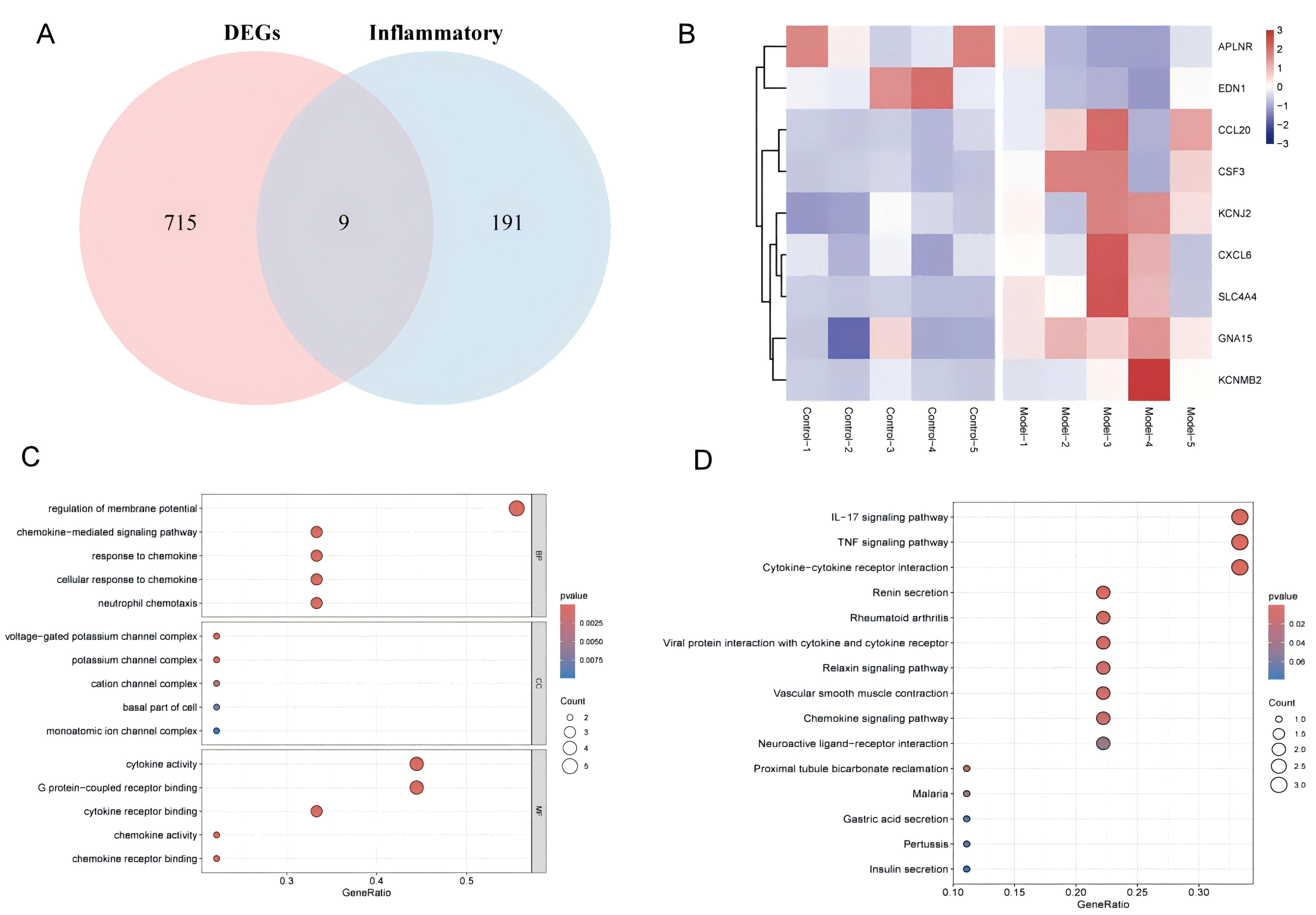
(A) The Venn diagram illustrated the intersection relationship between the DEGs in RNA-seq and the immune-related genes. (B) Heatmap of expression levels of candidate DEGs. (C) GO enrichment of the candidate DEGs. (D) KEGG enrichment of the candidate DEGs.
Figure 3. Correlation analysis of DEGs expression and immune infiltration.
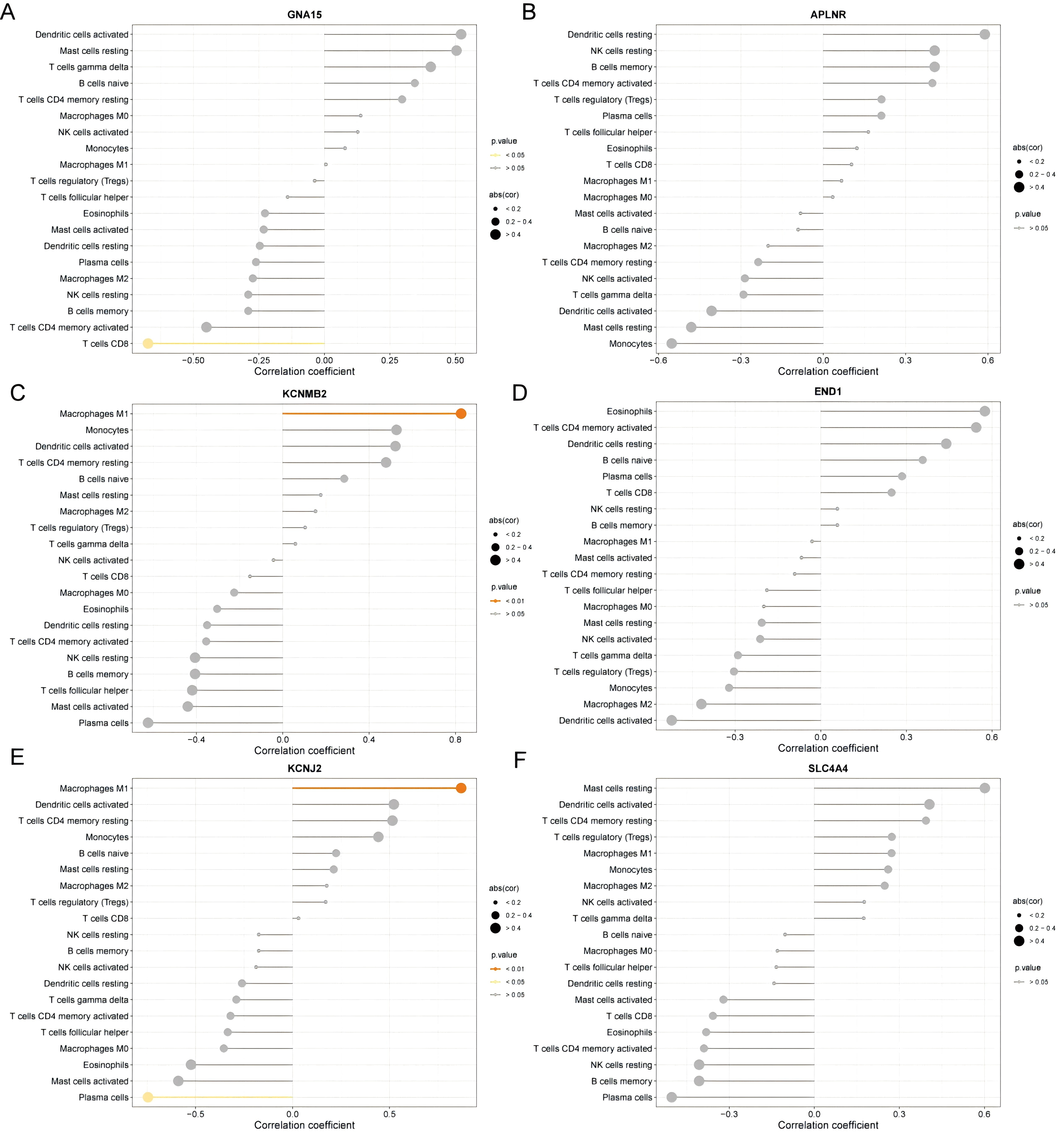
(A) GNA15; (B) APLNR; (C) KCNMB2; (D) END1; (E) KCNJ2; (F) SLC4A4. P<0.01.
Figure 4. Verify the expression levels of candidate DEGs in clinical samples.
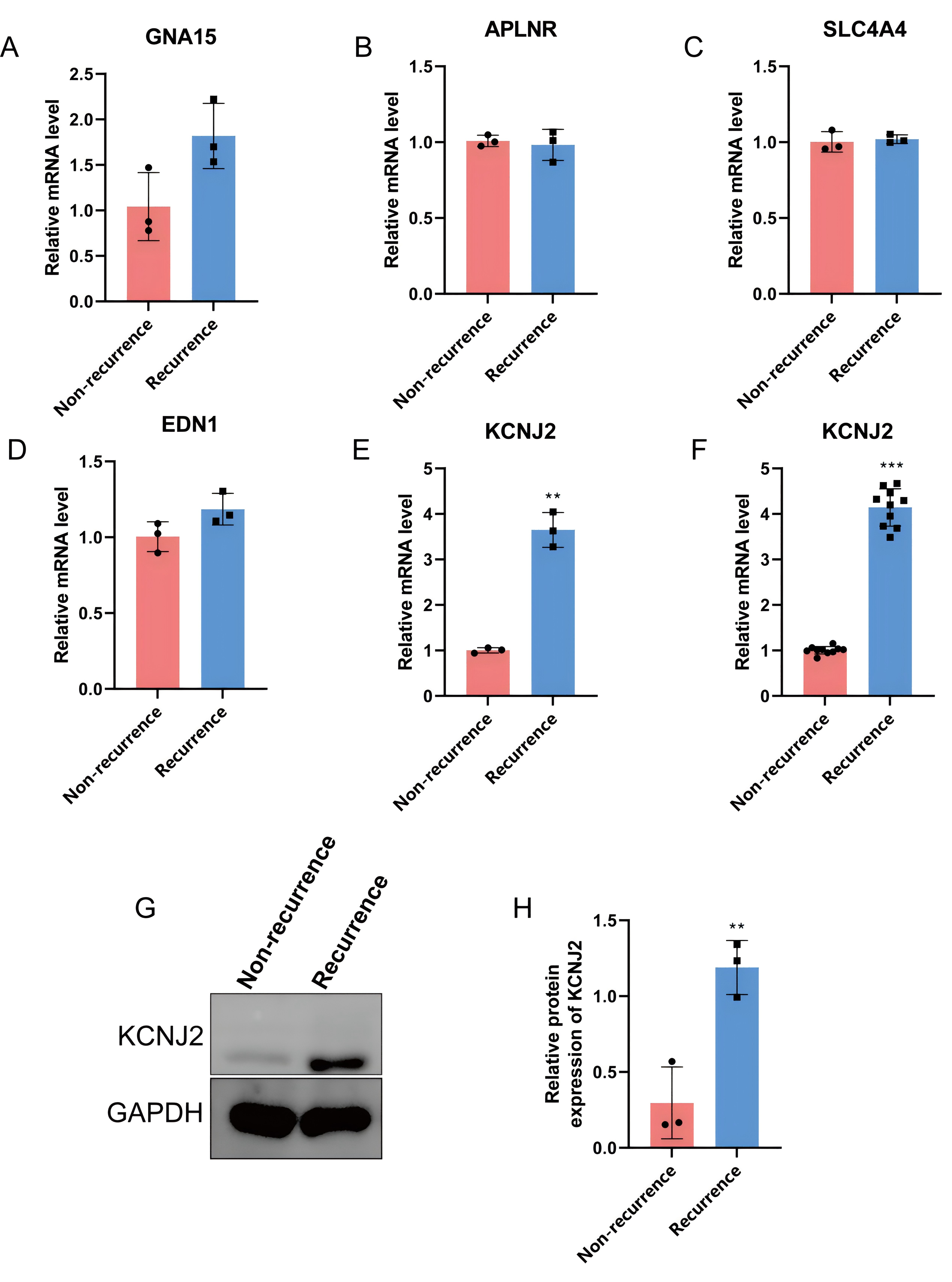
(A-E) qPCR was performed to assess the mRNA expression levels of DEGs, n=3 per group. (F) RT-qPCR was performed to assess KCNJ2 mRNA expression, n = 10 per group. (G-H) Western blotting was performed to assess the protein expression levels of KCNJ2, n=3 per group. Data are presented as mean ± SD, **p < 0.01, ***p < 0.001.
Macrophage M1 polarization associates with KCNJ2 expression in recurrent CRS
To clarify whether abnormal macrophage polarization occurs during the recurrence of chronic sinusitis after surgery and whether it is related to KCNJ2 expression, we evaluated the expression of M1 and M2 macrophage marker genes between the recurrence and non-recurrence groups. As shown in Figure 5, the mRNA levels of CD86 and iNOS were significantly elevated in the recurrence group, whereas the expression levels of CD163 and CD206 were markedly reduced. Notably, the expression of CD86 and iNOS was positively correlated with KCNJ2, while the expression of CD163, CD206, and KCNJ2 was negatively correlated.
We subsequently measured serum inflammatory cytokine levels using ELISA. Serum IL-6 and TNF-α levels were significantly higher in the recurrence group than in the non-recurrence group, whereas serum TGF-β levels were significantly lower (Figure 6A-C). Furthermore, tissue KCNJ2 expression was positively correlated with serum IL-6 and TNF-α levels but negatively correlated with serum TGF-β levels (Figure 6D-F). Collectively, these results demonstrate that elevated tissue KCNJ2 expression is associated with increased systemic pro-inflammatory cytokine levels and reduced TGF-β levels in patients with postoperative recurrence of CRS.
Figure 5. The mRNA expressions of macrophage M1- and M2-associated macrophage markers in different groups.
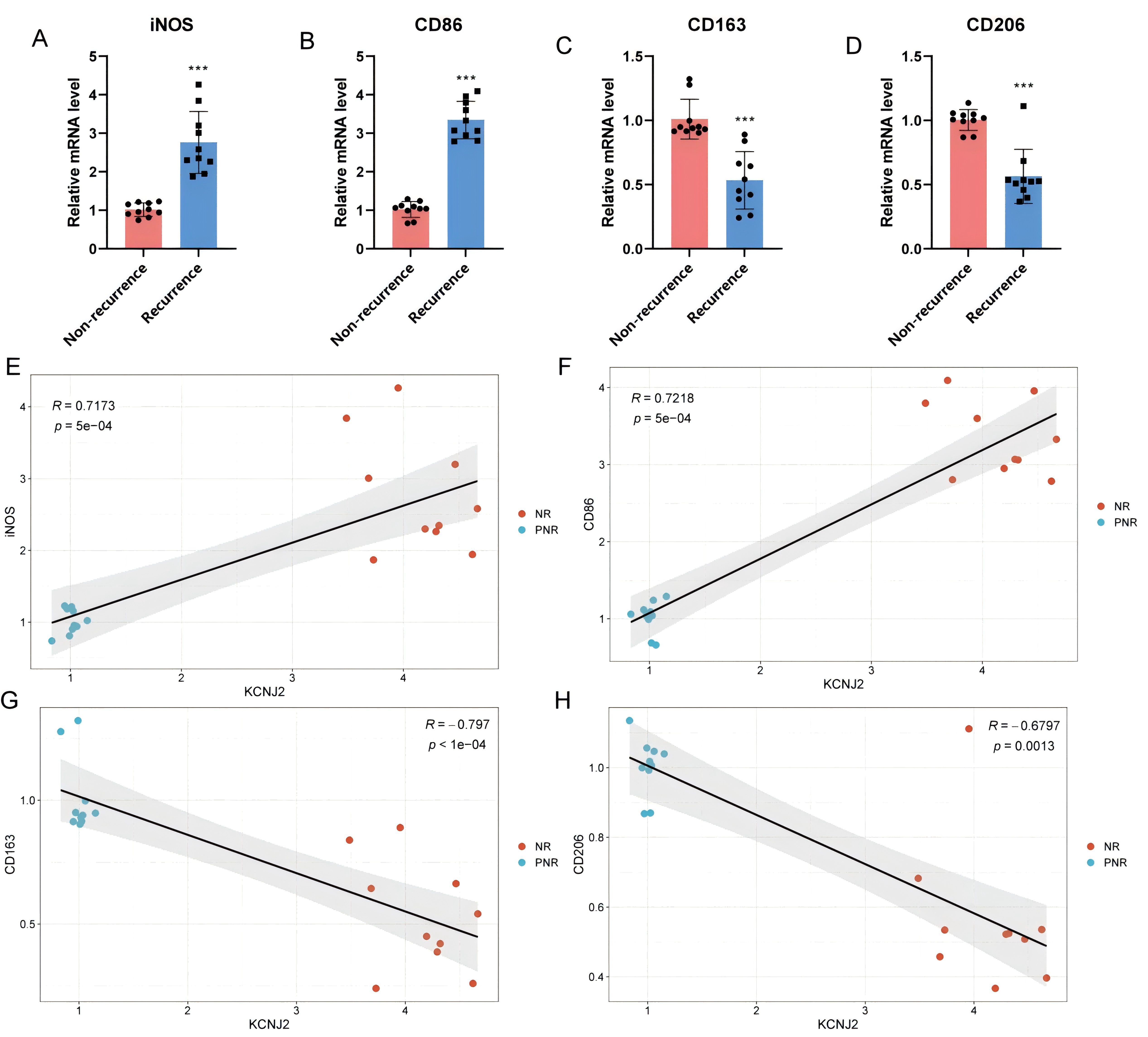
(A-D) qPCR was used to detect the expression of M1 and M2 macrophage marker genes. n = 10, per group. Data are presented as mean ± SD, ***P < 0.001. (E-H) Spearman correlation analysis of KCNJ2 expression with the marker genes, ***P < 0.001.
Figure 6. Altered serum cytokine levels in patients with postoperative recurrence of CRS.
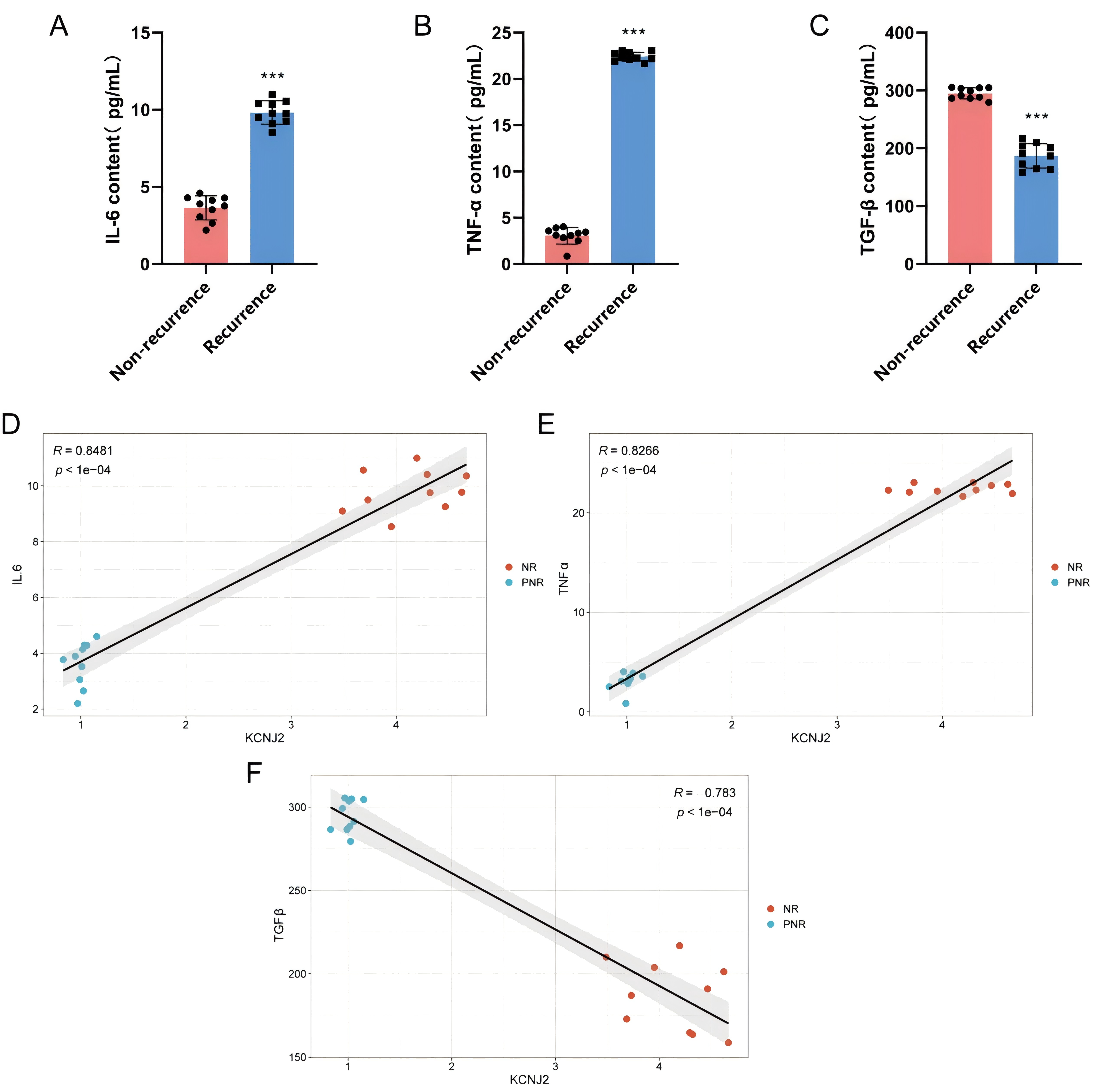
(A-C) ELISA was used to determine serum IL-6, TNF-α, and TGF-β levels in the non-recurrence and recurrence groups, with n = 10 independent patients per group. (D-F) Spearman correlation analysis between tissue KCNJ2 expression and serum cytokine levels. ***P < 0.001.
Discussion
CRS is a highly heterogeneous disease with a high postoperative recurrence rate. Despite extensive research on the pathogenesis of CRS and its subtypes, no predictive model for CRS postoperative recurrence has been established to date. Studies have demonstrated that persistent inflammatory responses and immune dysregulation may be the core factors driving postoperative recurrence. Therefore, from the perspective of immune regulation, screening key biomarkers associated with CRS postoperative recurrence is of great significance for the management of postoperative recurrence in patients, the reduction of recurrence rates, and the alleviation of burdens on patients and society.
RNA-seq, as one of the commonly used detection techniques, was often employed to identify disease markers. In this study, we sequenced the nasal sinus mucosal tissues of patients with postoperative recurrence and those without recurrence, and identified a total of 724 DEGs. These genes were enriched in immune inflammation-related pathways, suggesting that immune system dysregulation might play an important role in disease recurrence. Therefore, we downloaded immune inflammation-related gene sets from the database and performed intersection analysis with the sequenced differential gene set. A total of 9 DEGs were obtained. To further select the key genes, we combined it with immune infiltration analysis, and the results showed that only KCNJ2 was highly expressed in the disease and significantly positively correlated with M1 macrophages.
KCNJ2, as one of the members of the inward rectifying potassium ion channel subfamily J, is widely expressed in various cells such as neurons, skeletal muscles, cardiomyocytes, and immune cells [24–25]. Studies have shown that phosphatidylinositol-4,5-bisphosphate, by binding to KCNJ2, can open the inward potassium ion channel Kir2.1, thereby regulating the membrane potential [26], providing a theoretical basis for the participation of KCNJ2 in cell function regulation. In recent years, numerous reports have indicated that ion channels are closely related to the regulation of the mucosal barrier and inflammatory cells [27–28]. As one of the most widespread ion channels in the human body, potassium ion channels are widely expressed in various cells [24]. Lin et al. discovered that knocking down KCa3.1 could reduce the number of goblet cells and mast cells in the nasal mucosa of allergic rhinitis mice, as well as the levels of inflammatory cytokines IL-4, IL-9, and IL-17 in nasal lavage fluid [29]. The lack of KCa3.1 in T lymphocytes, B lymphocytes, mast cells, and bronchial smooth muscle cells has a protective effect against OVA-induced asthma [30], demonstrating the important role of potassium ion channels in regulating respiratory inflammatory diseases. The sequencing results of this study showed that the expression level of KCNJ2 in the sinus mucosa tissue of the recurrence group was significantly higher than that of the non-recurrence group. Combined with the regulatory role of potassium ion channels in respiratory inflammation, it suggests that KCNJ2 may be involved in the pathological process of sinusitis recurrence after surgery. Subsequently, to eliminate possible errors in the sequencing process, we collected 10 clinical samples for further verification. The results showed that the expression of KCNJ2 was significantly increased, consistent with the sequencing results.
The association between KCNJ2 and macrophage function has been documented previously. KCNJ2 is expressed in various macrophage populations, including microglia, bone marrow-derived macrophages, and THP-1 macrophages [31–32]. Yu et al. demonstrated that KCNJ2 is essential for maintaining the electrophysiological properties of macrophages and positively regulates macrophage inflammatory activation in vitro [33]. Furthermore, Chen et al. reported that knockdown of KCNJ2 markedly suppressed LPS-induced M1 macrophage polarization [34], suggesting that KCNJ2 contributes to M1 macrophage polarization.
In the present study, we observed that the expression of M1 macrophage markers iNOS and CD86 was significantly elevated in tissues from patients with postoperative recurrence, whereas the expression of M2 markers CD163 and CD206 was markedly reduced. Meanwhile, the levels of pro-inflammatory cytokines TNF-α and IL-6 were increased, while the anti-inflammatory factor TGF-β was decreased. These findings were consistent with previous reports [35], further supporting that aberrant M1 macrophage polarization may contribute to postoperative recurrence of chronic sinusitis. Notably, studies on postoperative recurrence of CRSwNP often reported a predominant M2 macrophage phenotype, which differs partially from our observations [36–37]. We speculate that this discrepancy may arise from distinct pathological processes between the two phenotypes. CRSwNP is characterized by nasal polyp formation, epithelial-mesenchymal transition (EMT), and nasal mucosal remodeling [38]. M2 macrophages not only mediate anti-inflammatory responses but also secrete cytokines that promote mucosal fibrosis, tissue remodeling, and EMT [39–41]. Thus, a certain level of M2 polarization is required to sustain these pathological features in CRSwNP. In contrast, the phenotype examined in this study was dominated by persistent local inflammatory activation, which favored a shift toward M1 macrophage polarization. Although our study identified concurrent upregulation of KCNJ2 and enhanced M1 macrophage polarization in patients with postoperative recurrence, the functional link between these two events remained unclear. We therefore performed correlation analysis. The results showed that KCNJ2 expression was significantly and positively correlated with M1 macrophage markers and pro-inflammatory cytokines TNFα and IL-6, but negatively correlated with M2 macrophage markers. Collectively, based on our findings and previous literature, we propose that KCNJ2 may promote M1 macrophage polarization and the subsequent release of pro-inflammatory mediators including TNFα and IL-6, thereby exacerbating local chronic inflammation in the nasal mucosa and contributing to the pathogenesis of postoperative recurrence of CRS.
Conclusion
This study shows that the transcriptional profiles of patients with postoperative recurrence are significantly different from those of patients without recurrence. Among these potential DEGs, KCNJ2 shows potential predictive ability for recurrence. The clinical sample validation results indicate that the expression of KCNJ2 in postoperative recurrence patients is significantly higher than that in non-recurrence patients. Moreover, KCNJ2 expression was elevated in patients with established postoperative recurrence and was associated with M1 macrophage polarization. These findings support KCNJ2 as a candidate recurrence-associated biomarker rather than a prospectively validated predictive marker.
Abbreviations
CRS: Chronic rhinosinusitis; CRSwNP: chronic sinusitis with nasal polyps; KCNJ2: potassium inwardly rectifying channel subfamily J member 2; RNA-seq: RNA sequencing; PCA: Principal Component Analysis; KEGG: Kyoto Encyclopedia of Genes and Genomes; DEGs: Differentially expressed genes; GO: Gene Ontology.
Declarations
Ethics approval and consent to participate
This study was approved by the Medical Ethics Committee of Bozhou People’s Hospital (Approval No. 2023-228). Written informed consent was obtained from all participants before sample collection. The study was conducted in accordance with the Declaration of Helsinki.
Acknowledgements
Not Applicable.
Consent for publication
All authors have reviewed and approved the final version of the manuscript and consent to its publication.
Availability of data and materials
The data generated or analyzed during this study are included in this article and its supplementary information files. Additional data are available from the corresponding author upon reasonable request.
Competing interests
The authors declare that they have no competing interests.
Funding
This work was supported by the Key Research and Development Project of Bozhou City in 2023 (bzzc2023062).
Authors’ contributions
M.Z. and W.C.: Study conception and design, data analysis and interpretation, and manuscript drafting. H.H. and C.F.: Experimental design, key experiments, data collection and analysis, and manuscript drafting and revision. M.H. and J.J.: Sample collection and processing, laboratory experiments, data organization, and result interpretation. G.D.: Study design participation, experimental assistance, and manuscript feedback. Q.J.: Data collection and organization, figure and table preparation, and manuscript revision. D.L.: Study design, data interpretation, manuscript revision, and research guidance. W.C. supervised the study, provided project administration, critically revised the manuscript, and approved the final version. All authors read and approved the final manuscript.
References
[1] Orlandi RR, Kingdom TT, Smith TL, Bleier BS, DeConde AS, Luong AU, et al. International consensus statement on allergy and rhinology: rhinosinusitis 2021. International Forum of Allergy & Rhinology. 2021;11(3):213-739. https://doi.org/10.1002/alr.22741
[2] Kato A, Peters AT, Stevens WW, Schleimer RP, Tan BK, Kern RC. Endotypes of chronic rhinosinusitis: relationships to disease phenotypes, pathogenesis, clinical findings, and treatment approaches. Allergy. 2022;77(3):812-826. https://doi.org/10.1111/all.15074
[3] Almosnino G, Little RE. Surgical management of rhinosinusitis for the allergist-immunologist. Annals of Allergy, Asthma & Immunology. 2023;131(3):311-316. https://doi.org/10.1016/j.anai.2023.05.015
[4] Maspero JF, Khan AH, Philpott C, Hellings PW, Hopkins C, Wagenmann M, et al. Health-related quality of life impairment among patients with severe chronic rhinosinusitis with nasal polyps in the SINUS-24 trial. Journal of Asthma and Allergy. 2023;16:323-332. https://doi.org/10.2147/JAA.S372598
[5] Khalmuratova R, Park JW, Shin HW. Immune cell responses and mucosal barrier disruptions in chronic rhinosinusitis. Immune Network. 2017;17(1):60-67. https://doi.org/10.4110/in.2017.17.1.60
[6] Kim JW, Hong SL, Kim YK, Lee CH, Min YG, Rhee CS. Histological and immunological features of non-eosinophilic nasal polyps. Otolaryngology-Head and Neck Surgery. 2007;137(6):925-930. https://doi.org/10.1016/j.otohns.2007.07.036
[7] Shi JB, Fu QL, Zhang H, Cheng L, Wang YJ, Zhu DD, et al. Epidemiology of chronic rhinosinusitis: results from a cross-sectional survey in seven Chinese cities. Allergy. 2015;70(5):533-539. https://doi.org/10.1111/all.12577
[8] Leopold DA, Elkayam D, Messina JC, Kosik-Gonzalez C, Djupesland PG, Mahmoud RA. NAVIGATE II: randomized, double-blind trial of the exhalation delivery system with fluticasone for nasal polyposis. Journal of Allergy and Clinical Immunology. 2019;143(1):126-134.e5. https://doi.org/10.1016/j.jaci.2018.06.010
[9] Brzost J, Czerwaty K, Dżaman K, Ludwig N, Piszczatowska K, Szczepański MJ. Perspectives in therapy of chronic rhinosinusitis. Diagnostics. 2022;12(10):2301. https://doi.org/10.3390/diagnostics12102301
[10] Grayson JW, Hopkins C, Mori E, Senior BA, Harvey RJ. Contemporary classification of chronic rhinosinusitis beyond polyps vs no polyps: a review. JAMA Otolaryngology-Head & Neck Surgery. 2020;146(9):831-838. https://doi.org/10.1001/jamaoto.2020.1453
[11] Bouatay R, Bouaziz N, Abdelali M, Zrig A, El Korbi A, Ferjaoui M, et al. Endoscopic sinus surgery for chronic rhino sinusitis with nasal polyps: predictive factors of recurrence. Ear, Nose & Throat Journal. 2024;Online ahead of print:1455613241295494. https://doi.org/10.1177/01455613241295494
[12] Liu X, Peng Y, Guo L, Xiong W, Liao W, Fan J. Unveiling and validating biomarkers related to the IL-10 family in chronic sinusitis with nasal polyps: insights from transcriptomics and single-cell RNA sequencing analysis. Frontiers in Molecular Biosciences. 2025;11:1513951. https://doi.org/10.3389/fmolb.2024.1513951
[13] Wang Y, Song X, Jin M, Lu J. Characterization of the immune microenvironment and identification of biomarkers in chronic rhinosinusitis with nasal polyps using single-cell RNA sequencing and transcriptome analysis. Journal of Inflammation Research. 2024;17:253-277. https://doi.org/10.2147/JIR.S440409
[14] Lv J, Jiang F. Integrative causal inference and predictive modeling reveal the iron-related gene SLC17A4 as a key biomarker in chronic rhinosinusitis. Frontiers in Immunology. 2026;16:1645726. https://doi.org/10.3389/fimmu.2025.1645726
[15] Fan HL, Han ZT, Gong XR, Wu YQ, Fu YJ, Zhu TM, et al. Macrophages in CRSwNP: do they deserve more attention? International Immunopharmacology. 2024;134:112236. https://doi.org/10.1016/j.intimp.2024.112236
[16] Jiang Z, Zhu L. Update on the role of alternatively activated macrophages in asthma. Journal of Asthma and Allergy. 2016;9:101-107. https://doi.org/10.2147/JAA.S104508
[17] Tarique AA, Logan J, Thomas E, Holt PG, Sly PD, Fantino E. Phenotypic, functional, and plasticity features of classical and alternatively activated human macrophages. American Journal of Respiratory Cell and Molecular Biology. 2015;53(5):676-688. https://doi.org/10.1165/rcmb.2015-0012OC
[18] Takabayashi T, Kato A, Peters AT, Hulse KE, Suh LA, Carter R, et al. Increased expression of factor XIII-A in patients with chronic rhinosinusitis with nasal polyps. Journal of Allergy and Clinical Immunology. 2013;132(3):584-592.e4. https://doi.org/10.1016/j.jaci.2013.02.003
[19] Wang K, Yu J, Luo Q, Bai Y, Gao Q, Yu R. siHIF-1α-loaded micellar nanoparticles inhibit M1 macrophage activation to ameliorate chronic rhinosinusitis. Free Radical Biology and Medicine. 2025;237:403-418. https://doi.org/10.1016/j.freeradbiomed.2025.05.433
[20] Kanehisa M, Furumichi M, Sato Y, Matsuura Y, Ishiguro-Watanabe M. KEGG: biological systems database as a model of the real world. Nucleic Acids Research. 2025;53(D1):D672-D677. https://doi.org/10.1093/nar/gkae909
[21] Zhuang M, Jiao J, Zhang Q, Pan S, Zhang Y, He T, et al. Ruxolitinib, a novel JAK/STAT-targeted therapy through restoring mucosal homeostasis for chronic rhinosinusitis with nasal polyps. Journal of Allergy and Clinical Immunology. 2025;156(5):1219-1233. https://doi.org/10.1016/j.jaci.2025.07.020
[22] Ayers CM, Schlosser RJ, O’Connell BP, Atkinson C, Mulligan RM, Casey SE, et al. Increased presence of dendritic cells and dendritic cell chemokines in the sinus mucosa of chronic rhinosinusitis with nasal polyps and allergic fungal rhinosinusitis. International Forum of Allergy & Rhinology. 2011;1(4):296-302. https://doi.org/10.1002/alr.20046
[23] Klingler AI, Stevens WW, Tan BK, Peters AT, Poposki JA, Grammer LC, et al. Mechanisms and biomarkers of inflammatory endotypes in chronic rhinosinusitis without nasal polyps. Journal of Allergy and Clinical Immunology. 2021;147(4):1306-1317. https://doi.org/10.1016/j.jaci.2020.11.037
[24] Hager NA, McAtee CK, Lesko MA, O’Donnell AF. Inwardly rectifying potassium channel Kir2.1 and its “Kir-ious” regulation by protein trafficking and roles in development and disease. Frontiers in Cell and Developmental Biology. 2022;9:796136. https://doi.org/10.3389/fcell.2021.796136
[25] Ozekin YH, Isner T, Bates EA. Ion channel contributions to morphological development: insights from the role of Kir2.1 in bone development. Frontiers in Molecular Neuroscience. 2020;13:99. https://doi.org/10.3389/fnmol.2020.00099
[26] Hansen SB, Tao X, MacKinnon R. Structural basis of PIP2 activation of the classical inward rectifier K+ channel Kir2.2. Nature. 2011;477(7365):495-498. https://doi.org/10.1038/nature10370
[27] Backaert W, Steelant B, Hellings PW, Talavera K, Van Gerven L. A TRiP through the roles of transient receptor potential cation channels in type 2 upper airway inflammation. Current Allergy and Asthma Reports. 2021;21(3):20. https://doi.org/10.1007/s11882-020-00981-x
[28] Deng Z, Zhao Y, Ma Z, Zhang M, Wang H, Yi Z, et al. Pathophysiological role of ion channels and transporters in gastrointestinal mucosal diseases. Cellular and Molecular Life Sciences. 2021;78(24):8109-8125. https://doi.org/10.1007/s00018-021-04011-5
[29] Lin H, Zheng C, Li J, Yang C, Hu L. Lentiviral shRNA against KCa3.1 inhibits allergic response in allergic rhinitis and suppresses mast cell activity via the PI3K/AKT signaling pathway. Scientific Reports. 2015;5:13127. https://doi.org/10.1038/srep13127
[30] Pelletier L, Savignac M. Involvement of ion channels in allergy. Current Opinion in Immunology. 2018;52:60-67. https://doi.org/10.1016/j.coi.2018.04.006
[31] Vicente R, Escalada A, Coma M, Fuster G, Sánchez-Tilló E, López-Iglesias C, et al. Differential voltage-dependent K+ channel responses during proliferation and activation in macrophages. Journal of Biological Chemistry. 2003;278(47):46307-46320. https://doi.org/10.1074/jbc.M304388200
[32] Qiao W, Wong KHM, Shen J, Wang W, Wu J, Li J, et al. TRPM7 kinase-mediated immunomodulation in macrophage plays a central role in magnesium ion-induced bone regeneration. Nature Communications. 2021;12(1):2885. https://doi.org/10.1038/s41467-021-23005-2
[33] Yu W, Wang Z, Yu X, Zhao Y, Xie Z, Zhang K, et al. Kir2.1-mediated membrane potential promotes nutrient acquisition and inflammation through regulation of nutrient transporters. Nature Communications. 2022;13(1):3544. https://doi.org/10.1038/s41467-022-31149-y
[34] Chen K, Man Q, Miao J, Xu W, Zheng Y, Zhou X, et al. Kir2.1 channel regulates macrophage polarization via the Ca2+/CaMKII/ERK/NF-κB signaling pathway. Journal of Cell Science. 2022;135(13):jcs259544. https://doi.org/10.1242/jcs.259544
[35] Wang T, Chen Y, Gao R, Shui J, Xie B. Overexpression of AXL on macrophages associates with disease severity and recurrence in chronic rhinosinusitis with nasal polyps. International Immunopharmacology. 2023;121:110449. https://doi.org/10.1016/j.intimp.2023.110449
[36] Zhong B, Du J, Liu F, Liu Y, Liu S, Zhang J, et al. Hypoxia-induced factor-1α induces NLRP3 expression by M1 macrophages in noneosinophilic chronic rhinosinusitis with nasal polyps. Allergy. 2021;76(2):582-586. https://doi.org/10.1111/all.14571
[37] Bao X, Liu B, Jiang Y, Feng T, Cao W, Shi J, et al. Loss of SENP3 mediated the formation of nasal polyps in nasal mucosal inflammation by increasing alternatively activated macrophages. Immunity, Inflammation and Disease. 2023;11(2):e781. https://doi.org/10.1002/iid3.781
[38] Dong Z, Jiang Y, Zhang J, Zheng C, Chen H, Tang W, et al. FERMT1 contributes to the epithelial-mesenchymal transition of chronic rhinosinusitis with nasal polyps via PI3K/Akt signaling. International Immunopharmacology. 2024;143(Pt 3):113545. https://doi.org/10.1016/j.intimp.2024.113545
[39] Lin H, Lin D, Xiong XS, Dai XX, Lin T. Role of platelet-derived growth factor-α in eosinophilic and non-eosinophilic chronic rhinosinusitis with nasal polyps. International Forum of Allergy & Rhinology. 2014;4(11):909-914. https://doi.org/10.1002/alr.21419
[40] Van Bruaene N, Derycke L, Perez-Novo CA, Gevaert P, Holtappels G, De Ruyck N, et al. TGF-β signaling and collagen deposition in chronic rhinosinusitis. Journal of Allergy and Clinical Immunology. 2009;124(2):253-259.e2. https://doi.org/10.1016/j.jaci.2009.04.013
[41] Mantovani A, Biswas SK, Galdiero MR, Sica A, Locati M. Macrophage plasticity and polarization in tissue repair and remodelling. Journal of Pathology. 2013;229(2):176-185. https://doi.org/10.1002/path.4133
 Figures
 References
 Peer
 Information
Figure 1. RNA-seq identified a distinctive gene profile in CRS patients between the Recurrence and Non-recurrence groups.
(A) PCA model of the transcriptomics data between the two groups. (B) Volcano plot showing genes upregulated and downregulated by RNA-seq. (C) Bar chart shows the number of DEGs between the two groups. (D) GO enrichment analysis. (E) KEGG enrichment analysis. Control, Non-recurrence; Model, Recurrence. n=5.
Figure 2. Bioinformatics analysis identified 9 immune-related DEGs.
(A) The Venn diagram illustrated the intersection relationship between the DEGs in RNA-seq and the immune-related genes. (B) Heatmap of expression levels of candidate DEGs. (C) GO enrichment of the candidate DEGs. (D) KEGG enrichment of the candidate DEGs.
Figure 3. Correlation analysis of DEGs expression and immune infiltration.
(A) GNA15; (B) APLNR; (C) KCNMB2; (D) END1; (E) KCNJ2; (F) SLC4A4. P<0.01.
Figure 4. Verify the expression levels of candidate DEGs in clinical samples.
(A-E) qPCR was performed to assess the mRNA expression levels of DEGs, n=3 per group. (F) RT-qPCR was performed to assess KCNJ2 mRNA expression, n = 10 per group. (G-H) Western blotting was performed to assess the protein expression levels of KCNJ2, n=3 per group. Data are presented as mean ± SD, **p < 0.01, ***p < 0.001.
Figure 5. The mRNA expressions of macrophage M1- and M2-associated macrophage markers in different groups.
(A-D) qPCR was used to detect the expression of M1 and M2 macrophage marker genes. n = 10, per group. Data are presented as mean ± SD, ***P < 0.001. (E-H) Spearman correlation analysis of KCNJ2 expression with the marker genes, ***P < 0.001.
Figure 6. Altered serum cytokine levels in patients with postoperative recurrence of CRS.
(A-C) ELISA was used to determine serum IL-6, TNF-α, and TGF-β levels in the non-recurrence and recurrence groups, with n = 10 independent patients per group. (D-F) Spearman correlation analysis between tissue KCNJ2 expression and serum cytokine levels. ***P < 0.001.
[1] Orlandi RR, Kingdom TT, Smith TL, Bleier BS, DeConde AS, Luong AU, et al. International consensus statement on allergy and rhinology: rhinosinusitis 2021. International Forum of Allergy & Rhinology. 2021;11(3):213-739. https://doi.org/10.1002/alr.22741
[2] Kato A, Peters AT, Stevens WW, Schleimer RP, Tan BK, Kern RC. Endotypes of chronic rhinosinusitis: relationships to disease phenotypes, pathogenesis, clinical findings, and treatment approaches. Allergy. 2022;77(3):812-826. https://doi.org/10.1111/all.15074
[3] Almosnino G, Little RE. Surgical management of rhinosinusitis for the allergist-immunologist. Annals of Allergy, Asthma & Immunology. 2023;131(3):311-316. https://doi.org/10.1016/j.anai.2023.05.015
[4] Maspero JF, Khan AH, Philpott C, Hellings PW, Hopkins C, Wagenmann M, et al. Health-related quality of life impairment among patients with severe chronic rhinosinusitis with nasal polyps in the SINUS-24 trial. Journal of Asthma and Allergy. 2023;16:323-332. https://doi.org/10.2147/JAA.S372598
[5] Khalmuratova R, Park JW, Shin HW. Immune cell responses and mucosal barrier disruptions in chronic rhinosinusitis. Immune Network. 2017;17(1):60-67. https://doi.org/10.4110/in.2017.17.1.60
[6] Kim JW, Hong SL, Kim YK, Lee CH, Min YG, Rhee CS. Histological and immunological features of non-eosinophilic nasal polyps. Otolaryngology-Head and Neck Surgery. 2007;137(6):925-930. https://doi.org/10.1016/j.otohns.2007.07.036
[7] Shi JB, Fu QL, Zhang H, Cheng L, Wang YJ, Zhu DD, et al. Epidemiology of chronic rhinosinusitis: results from a cross-sectional survey in seven Chinese cities. Allergy. 2015;70(5):533-539. https://doi.org/10.1111/all.12577
[8] Leopold DA, Elkayam D, Messina JC, Kosik-Gonzalez C, Djupesland PG, Mahmoud RA. NAVIGATE II: randomized, double-blind trial of the exhalation delivery system with fluticasone for nasal polyposis. Journal of Allergy and Clinical Immunology. 2019;143(1):126-134.e5. https://doi.org/10.1016/j.jaci.2018.06.010
[9] Brzost J, Czerwaty K, Dżaman K, Ludwig N, Piszczatowska K, Szczepański MJ. Perspectives in therapy of chronic rhinosinusitis. Diagnostics. 2022;12(10):2301. https://doi.org/10.3390/diagnostics12102301
[10] Grayson JW, Hopkins C, Mori E, Senior BA, Harvey RJ. Contemporary classification of chronic rhinosinusitis beyond polyps vs no polyps: a review. JAMA Otolaryngology-Head & Neck Surgery. 2020;146(9):831-838. https://doi.org/10.1001/jamaoto.2020.1453
[11] Bouatay R, Bouaziz N, Abdelali M, Zrig A, El Korbi A, Ferjaoui M, et al. Endoscopic sinus surgery for chronic rhino sinusitis with nasal polyps: predictive factors of recurrence. Ear, Nose & Throat Journal. 2024;Online ahead of print:1455613241295494. https://doi.org/10.1177/01455613241295494
[12] Liu X, Peng Y, Guo L, Xiong W, Liao W, Fan J. Unveiling and validating biomarkers related to the IL-10 family in chronic sinusitis with nasal polyps: insights from transcriptomics and single-cell RNA sequencing analysis. Frontiers in Molecular Biosciences. 2025;11:1513951. https://doi.org/10.3389/fmolb.2024.1513951
[13] Wang Y, Song X, Jin M, Lu J. Characterization of the immune microenvironment and identification of biomarkers in chronic rhinosinusitis with nasal polyps using single-cell RNA sequencing and transcriptome analysis. Journal of Inflammation Research. 2024;17:253-277. https://doi.org/10.2147/JIR.S440409
[14] Lv J, Jiang F. Integrative causal inference and predictive modeling reveal the iron-related gene SLC17A4 as a key biomarker in chronic rhinosinusitis. Frontiers in Immunology. 2026;16:1645726. https://doi.org/10.3389/fimmu.2025.1645726
[15] Fan HL, Han ZT, Gong XR, Wu YQ, Fu YJ, Zhu TM, et al. Macrophages in CRSwNP: do they deserve more attention? International Immunopharmacology. 2024;134:112236. https://doi.org/10.1016/j.intimp.2024.112236
[16] Jiang Z, Zhu L. Update on the role of alternatively activated macrophages in asthma. Journal of Asthma and Allergy. 2016;9:101-107. https://doi.org/10.2147/JAA.S104508
[17] Tarique AA, Logan J, Thomas E, Holt PG, Sly PD, Fantino E. Phenotypic, functional, and plasticity features of classical and alternatively activated human macrophages. American Journal of Respiratory Cell and Molecular Biology. 2015;53(5):676-688. https://doi.org/10.1165/rcmb.2015-0012OC
[18] Takabayashi T, Kato A, Peters AT, Hulse KE, Suh LA, Carter R, et al. Increased expression of factor XIII-A in patients with chronic rhinosinusitis with nasal polyps. Journal of Allergy and Clinical Immunology. 2013;132(3):584-592.e4. https://doi.org/10.1016/j.jaci.2013.02.003
[19] Wang K, Yu J, Luo Q, Bai Y, Gao Q, Yu R. siHIF-1α-loaded micellar nanoparticles inhibit M1 macrophage activation to ameliorate chronic rhinosinusitis. Free Radical Biology and Medicine. 2025;237:403-418. https://doi.org/10.1016/j.freeradbiomed.2025.05.433
[20] Kanehisa M, Furumichi M, Sato Y, Matsuura Y, Ishiguro-Watanabe M. KEGG: biological systems database as a model of the real world. Nucleic Acids Research. 2025;53(D1):D672-D677. https://doi.org/10.1093/nar/gkae909
[21] Zhuang M, Jiao J, Zhang Q, Pan S, Zhang Y, He T, et al. Ruxolitinib, a novel JAK/STAT-targeted therapy through restoring mucosal homeostasis for chronic rhinosinusitis with nasal polyps. Journal of Allergy and Clinical Immunology. 2025;156(5):1219-1233. https://doi.org/10.1016/j.jaci.2025.07.020
[22] Ayers CM, Schlosser RJ, O’Connell BP, Atkinson C, Mulligan RM, Casey SE, et al. Increased presence of dendritic cells and dendritic cell chemokines in the sinus mucosa of chronic rhinosinusitis with nasal polyps and allergic fungal rhinosinusitis. International Forum of Allergy & Rhinology. 2011;1(4):296-302. https://doi.org/10.1002/alr.20046
[23] Klingler AI, Stevens WW, Tan BK, Peters AT, Poposki JA, Grammer LC, et al. Mechanisms and biomarkers of inflammatory endotypes in chronic rhinosinusitis without nasal polyps. Journal of Allergy and Clinical Immunology. 2021;147(4):1306-1317. https://doi.org/10.1016/j.jaci.2020.11.037
[24] Hager NA, McAtee CK, Lesko MA, O’Donnell AF. Inwardly rectifying potassium channel Kir2.1 and its “Kir-ious” regulation by protein trafficking and roles in development and disease. Frontiers in Cell and Developmental Biology. 2022;9:796136. https://doi.org/10.3389/fcell.2021.796136
[25] Ozekin YH, Isner T, Bates EA. Ion channel contributions to morphological development: insights from the role of Kir2.1 in bone development. Frontiers in Molecular Neuroscience. 2020;13:99. https://doi.org/10.3389/fnmol.2020.00099
[26] Hansen SB, Tao X, MacKinnon R. Structural basis of PIP2 activation of the classical inward rectifier K+ channel Kir2.2. Nature. 2011;477(7365):495-498. https://doi.org/10.1038/nature10370
[27] Backaert W, Steelant B, Hellings PW, Talavera K, Van Gerven L. A TRiP through the roles of transient receptor potential cation channels in type 2 upper airway inflammation. Current Allergy and Asthma Reports. 2021;21(3):20. https://doi.org/10.1007/s11882-020-00981-x
[28] Deng Z, Zhao Y, Ma Z, Zhang M, Wang H, Yi Z, et al. Pathophysiological role of ion channels and transporters in gastrointestinal mucosal diseases. Cellular and Molecular Life Sciences. 2021;78(24):8109-8125. https://doi.org/10.1007/s00018-021-04011-5
[29] Lin H, Zheng C, Li J, Yang C, Hu L. Lentiviral shRNA against KCa3.1 inhibits allergic response in allergic rhinitis and suppresses mast cell activity via the PI3K/AKT signaling pathway. Scientific Reports. 2015;5:13127. https://doi.org/10.1038/srep13127
[30] Pelletier L, Savignac M. Involvement of ion channels in allergy. Current Opinion in Immunology. 2018;52:60-67. https://doi.org/10.1016/j.coi.2018.04.006
[31] Vicente R, Escalada A, Coma M, Fuster G, Sánchez-Tilló E, López-Iglesias C, et al. Differential voltage-dependent K+ channel responses during proliferation and activation in macrophages. Journal of Biological Chemistry. 2003;278(47):46307-46320. https://doi.org/10.1074/jbc.M304388200
[32] Qiao W, Wong KHM, Shen J, Wang W, Wu J, Li J, et al. TRPM7 kinase-mediated immunomodulation in macrophage plays a central role in magnesium ion-induced bone regeneration. Nature Communications. 2021;12(1):2885. https://doi.org/10.1038/s41467-021-23005-2
[33] Yu W, Wang Z, Yu X, Zhao Y, Xie Z, Zhang K, et al. Kir2.1-mediated membrane potential promotes nutrient acquisition and inflammation through regulation of nutrient transporters. Nature Communications. 2022;13(1):3544. https://doi.org/10.1038/s41467-022-31149-y
[34] Chen K, Man Q, Miao J, Xu W, Zheng Y, Zhou X, et al. Kir2.1 channel regulates macrophage polarization via the Ca2+/CaMKII/ERK/NF-κB signaling pathway. Journal of Cell Science. 2022;135(13):jcs259544. https://doi.org/10.1242/jcs.259544
[35] Wang T, Chen Y, Gao R, Shui J, Xie B. Overexpression of AXL on macrophages associates with disease severity and recurrence in chronic rhinosinusitis with nasal polyps. International Immunopharmacology. 2023;121:110449. https://doi.org/10.1016/j.intimp.2023.110449
[36] Zhong B, Du J, Liu F, Liu Y, Liu S, Zhang J, et al. Hypoxia-induced factor-1α induces NLRP3 expression by M1 macrophages in noneosinophilic chronic rhinosinusitis with nasal polyps. Allergy. 2021;76(2):582-586. https://doi.org/10.1111/all.14571
[37] Bao X, Liu B, Jiang Y, Feng T, Cao W, Shi J, et al. Loss of SENP3 mediated the formation of nasal polyps in nasal mucosal inflammation by increasing alternatively activated macrophages. Immunity, Inflammation and Disease. 2023;11(2):e781. https://doi.org/10.1002/iid3.781
[38] Dong Z, Jiang Y, Zhang J, Zheng C, Chen H, Tang W, et al. FERMT1 contributes to the epithelial-mesenchymal transition of chronic rhinosinusitis with nasal polyps via PI3K/Akt signaling. International Immunopharmacology. 2024;143(Pt 3):113545. https://doi.org/10.1016/j.intimp.2024.113545
[39] Lin H, Lin D, Xiong XS, Dai XX, Lin T. Role of platelet-derived growth factor-α in eosinophilic and non-eosinophilic chronic rhinosinusitis with nasal polyps. International Forum of Allergy & Rhinology. 2014;4(11):909-914. https://doi.org/10.1002/alr.21419
[40] Van Bruaene N, Derycke L, Perez-Novo CA, Gevaert P, Holtappels G, De Ruyck N, et al. TGF-β signaling and collagen deposition in chronic rhinosinusitis. Journal of Allergy and Clinical Immunology. 2009;124(2):253-259.e2. https://doi.org/10.1016/j.jaci.2009.04.013
[41] Mantovani A, Biswas SK, Galdiero MR, Sica A, Locati M. Macrophage plasticity and polarization in tissue repair and remodelling. Journal of Pathology. 2013;229(2):176-185. https://doi.org/10.1002/path.4133
Peer-review Terminology
Identity transparency: Single-anonymous
Reviewer interacts with: Editor
Details
This is an open access article under the terms of the Creative Commons Attribution License(http://creativecommons.org/licenses/by/4.0/), which permits use, distribution and reproduction in any medium, provided the original work is properly cited.
Publication History
Received 2026-05-26
Accepted 2026-07-15
Published 2026-07-30